
BACKGROUND
Rehabilitation of cleft lip and palate patients is a major challenge for Implantology since these patients require bone grafting procedures not only to achieve sufficient bone support for the installation of osseointegrable implants, but also to obtain a bone height suitable for the aesthetics of the prosthesis6,10,15,18,19,23,24.
Bone grafting is the procedure of choice for the closure of oronasal fistula3,4,6,8,12,14,18,19,25 and thereby allows dental eruption at the site, provides bone support for adjacent dental elements, stabilizes the premaxilla and creates support for the alar base6,18,19.
The rehabilitation of these patients through conventional prostheses presents functional, aesthetic and social problems2,6,10,19,23. In addition to circumventing these factors, rehabilitation with dental implants in these areas promotes bone stimulation, preventing reabsorption of the graft and ensuring the longevity of the benefits2,6,18,23.
With the introduction of dental implants, the qualification of the professionals and the biotechnologies involved, the indications for oral rehabilitation were expanded, modifying the treatment in several presentations of partial or total edentulism2,6,18,19. Thus, dental problems that, historically, were the most difficult to solve, can now be solved more efficiently2,6.
Cleft palate and cleft lip are among the most frequent congenital malformations, with an incidence of 1/10003,4,6,8,11,12,14,25. Approximately 75% of cases include a defect in the maxillary alveolar bone, and bone grafting, which is the therapy of choice, is necessary in 30% to 40% of patients3,4,6,11.
Regarding the classification of the various types of clefts, Kernahan et al. (1958) identified three main groups: I) Primary cleft palate - lip and alveolar ridge; II) Secondary palate clefts - hard palate and soft palate; III) Primary and secondary palate clefts - lip, dental alveolus and hard and soft palate.
Another classification, one of the most commonly used, is by Spina et al. (1972), modified, because it is simple, objective and practical enough to facilitate communication between professionals of a multidisciplinary team. This classification comprises four different groups and has as an anatomical reference point the incisive foramen that separates the primary palate from the secondary palate. I) Incisive pre-foramen clefts: Unilateral (incomplete or complete); Bilateral (incomplete or complete); Median (incomplete or complete). II) Incisive trans-foramen clefts: Unilateral; Bilateral; Median. III) Incised post-foramen clefts: Incomplete; or Complete. IV) Rare face clefts. It is necessary to take into account that the pre-foramen are related to the structure of the lips and alveoli, and that the trans-foramen and post-foramen are related to the hard and soft palate.
If the grafting procedure is performed before the canine eruption, orthodontic movement allows complete closure in 90% of the cases, and only 10% of prosthetic reconstruction is necessary. If the grafting occurs after the canine eruption, the possibility of closure by orthodontic mechanics is reduced to 72%, increasing prosthetic demand4,6,20.
Because of this indication of orthodontic therapy, as to the choice of materials for grafting, bone substitutes such as hydroxyapatite and lyophilized bone are not considered adequate, since they do not allow dental movement, either natural (eruption) or orthodontic1,6,7,10,20,22.
Cleft lip and palate patients often present agenesis, especially of lateral incisors10,17. Therefore, the importance of orthodontic intervention is highlighted, promoting the closure of the diastema by the mesialization of the tooth subsequent to the failure. In patients with agenesis of more than one tooth, this exclusively orthodontic closure is practically impossible, leading to the need for prosthetic rehabilitation 10,20.
The most frequent method of rehabilitation is fixed and removable partial dentures6,18,19,23. However, because they require wear of healthy dental elements and present functional, aesthetic and/or social difficulties, another therapeutic option was necessary for the effective resolution of these cases. The great success rate of osseointegrated implants has provided this new rehabilitation possibility, since a correct preparation of the receptor site has been performed through correct bone grafting techniques and after the end of the orofacial region2,6,18,23.
Three factors should be analyzed when performing bone grafting as a preparation of the implant site for implants4,6: 1) Extension of the bone defect - the bone defect may be incomplete or complete (if it presents oronasal fistula); 2) Adjacent soft tissues - always present low quality, result of the congenital defect, hypoplasia and scar fibrosis; 3) Fill material - it should be considered that two materials must be combined: one that fills the bone defect and another that serves as a framework for the first.
Although there are several types of materials for grafting, the one of choice is the autogenous bone, because it provides osteogenic cells (important in the first stage of bone formation) and does not activate the immune1,5,7,15,16,22,23,24. response The main disadvantages of autogenous bone are the need for another surgical site, increasing the morbidity of the procedure as a whole, and the availability of material from donor sites1,5,7,15,23.
Thus, the search for a viable alternative to the autogenous bone led to the homologous bone (allogeneic). Despite reducing surgical morbidity, the main disadvantage of the homologous bone is that it does not have osteogenic cells (lost during the preparation process, so as not to activate the immune response), not participating in the first stage of osteo-formation. Its contribution occurs only in the second phase of osteogenesis (osteoinduction) being purely passive, serving only as a framework1,5,7,10,15,16,23,24.
The present article aimed to report the procedure of bone grafting in a cleft lip and palate patient, aiming at the correction of the recipient bed for posterior rehabilitation with dental implant.
METHODS
A 24-year-old cleft lip and palate male patient (Fig. 1A, 1B) presented the clinical course of a postgraduate course in Implant Dentistry for rehabilitation with an osseointegrable implant. He had undergone previous orthodontic treatment for 26 months. The patient presented with deciduous elements 52 and 53 and agenesis of 12. A computed tomography (CT) scan of the region revealed a severe maxillary bone defect in the region of agenesis (Fig. 1C), with communication of the bone defect with the incisor canal (Fig. 1D, 1E), however, without oronasal fistula (unilateral pre-foramen cleft).
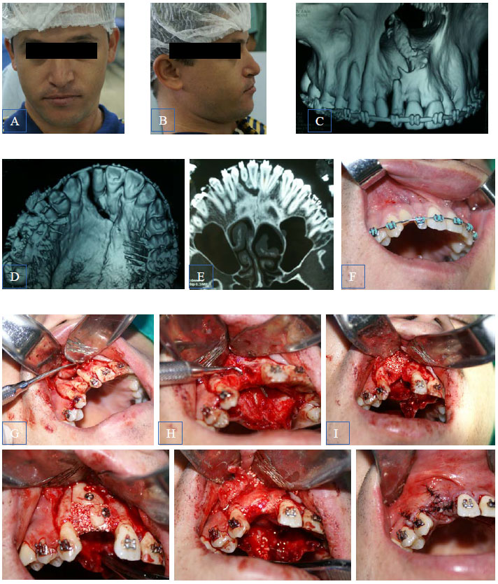 Fig.1 Procedure of homologous grafting in a cleft lip and palate patient. A, Front view; B, Side view; C, CT - front view; D, CT - occlusal view; E, CT - cut demonstrating communication of the bone defect with the incisor canal; F, intraoral view; G, detachment and exposure of the receptor site. Presence of deciduous elements 52-23 and agenesis of element 22; H, Probe demonstrating the communication of the bone defect with the incisor canal; I, Filling with inorganic bovine lyophilized bone; J, Fixation of the two blocks of homologous bone; K, Adaptation of the collagen membrane; L, Post-suture closure.
Based on the data from the complementary exams, surgical planning was performed to correct the implantation site. The proposal presented to the patient included the use of autogenous bone graft (obtained from the region of mandibular angle) and inorganic lyophilized bone. However, according to the patient's objection, the use of homologous bone and inorganic lyophilized bone was chosen. Despite the differences between the properties of these grafting options, there would be no compromise of the final result, since there would be no more orthodontic movement at the site.
RESULTS
After intrasulcular incision with total mucoperiosteal flap (Fig. 1G), the deciduous elements and curettage of the bone defect were removed, in order to remove any remaining tissues internally adhered to the defect. Secondly, the incisor canal was emptied and filled with inorganic bovine lyophilized bone (Fig. 1H, 1I) GEN-OX® (BAUMER). The maxillary defect was filled with homologous cortical bone of patella (from Bone Bank of Marília) and inorganic lyophilized bone GEN-OX® (BAUMER), the blocks were fixed by graft fixation screws (Fig. 1J) of 12 Mm and 10 mm (inches). The graft complex was covered by a GEN-DERM® collagen membrane (BAUMER) to protect the area (Fig. 1K). The surgical site was then closed without tension in order to avoid exposure during healing, with simple and horizontal and vertical mesodermal sutures (Fig. 1L).
DISCUSSION
Although several studies on bone grafting have shown excellent histological, radiographic and clinical results1,6,15,18 few studies have examined these results in grafts performed in cleft lip and palate patients10,13. An important difficulty in these patients is the fixation of the bone graft, since the instability may promote its reabsorption5,6,7,15. This leads to insufficient alveolar bone height and a large volume of graft becomes necessary1,10,15,18. In addition, frequently, acquired bone height is insufficient, requiring new grafting10,15.
Another difficulty is the loss of elasticity of the adjacent soft tissue, due to the healing fibrosing of other surgeries on site4,6,18 which leads to a great difficulty of closing by first intention. Due to its biological properties, the gold standard grafting material is the autologous bone, allowing the continuity of all the local physiological processes4,5,6,7,15,22,23,24. However, studies have shown a high rate of resorption in this region of bone clefts. The reason for this is attributed to the probable compression resulting from the upper lip and movement during phonation4.
Despite its physiological limitations1,5,7,15,16,23,24 homologous bone presented a high success rate in these patients, even presenting a lower level of resorption when compared to autologous bones1,5,7,15,24, but the mechanisms responsible for such differences are still unknown.
There are several products on the market designed to replace human bone (autologous or homologous). Each professional should select the best product, according to its characteristics and studies performed on it, for each clinical case1,7,10,15,20,22.
After years of research, as no substance with properties equivalent to autogenous bone was found, studies are have been performed for a better understanding of the process of incorporation of the grafts, in order to develop an ideal substitute, thus reducing surgical morbidity for the patient1,5.7,15,16,22,24.
Many studies report the experiences with osseointegratable implants in patients with fissures6,10,18,19,24. However, in most studies the sample was small and the follow-up period was relatively short10,18. Because of these limitations, potential risk factors for implant loss in these patients are difficult to determine. In addition, there is a lack of studies comparing the success rate of implants in both cleft lip and palate and non-cleft lip and palate patients10.
CONCLUSION
The grafting procedures in cleft lip and palate patients, aiming at preparing the receptor site for subsequent installation of osseointegrable implants, present a high rate of surgical success, providing strong bases for a full oral rehabilitation, from the functional, aesthetic and social points of view, promoting greater patient satisfaction.
Grafts with homologous bone for these situations can be performed safely and with significantly success rate, since correctly employed according to the type of rehabilitation treatment plan proposed by the professional.
REFERENCES
1. Baptista DA, Sorrilha A, Tormes TAM, Abdcune YA, Croci AT, Camargo OP et al. Estudo histológico dos enxertos ósseos homólogos humanos. Acta Ortop Bras 2003; 11: 220-4.
2. Block MS, Baughman DG. Reconstruction of severe anterior maxillary defects using distraction osteogenesis, bone grafts, and implants. J Oral Maxillofac Surg 2005; 63:291-7.
3. Corbo M, Dujardin T, de Maertelaer V, Malevez C, Glineur R. Dentocraniofacial morphology of 21 patients with unilateral cleft lip and palate: a cephalometric study. Cleft Palate Craniofac J. 2005; 42: 618-24.
4. Duskova M, Kotova M, Sedlackova K, Leamerova E, Horak J. Bone reconstruction of the maxillary alveolus for subsequent insertion of a dental implant in patients with cleft lip and palate. J Craniofac Surg. 2007; 18: 630-8.
5. Ellis III E, Sinn DP. Use of homologous bone in maxillofacial surgery. J Oral Maxillolac Surg 1993; 51: 1181-93.
6. Genden EM. Reconstruction of the mandible and the maxilla: the evolution of surgical technique. Arch Facial Plast Surg 2010; 12: 87-90.
7. Goiato MC, Anchieta RB, Pita MS, Santos DM. Reconstruction of skull defects: currently available materials. J Craniofac Surg 2009; 20: 1512-1518.
8. Honda Y, Suzuki A, Nakamura N, Ohishi M. Relationship between primary palatal form and maxillofacial growth in Japanese children with unilateral cleft lip and palate: infancy to adolescence. Cleft Palate Craniofac J 2002; 39:527-34.
9. Kernahan DA, Stark RB. A new classification for cleft lip and palate. Plast Reconstr Surg Transplant Bull 1958; 22: 435-41.
10. Kramer FJ, Baethqe C, Swennen G, Bremer B, Schwestka-Polly R, Dempf R. Dental implants in patients with orofacial clefts: a long-term follow-up study. Int J Oral Maxillofac. Surg 2005; 34: 715–21.
11. Liao YF, Mars M. Long-term effects of clefts on craniofacial morphology in patients with unilateral cleft lip and palate. Cleft Palate Craniofac J 2005; 42: 601-9.
12. Liao YF, Mars M. Long-term effects of palate repair on craniofacial morphology in patients with unilateral cleft lip and palate. Cleft Palate Craniofac J 2005; 42: 594-600.
13. Mulliken JB, Wu JK, Padwa BL. Repair of bilateral cleft lip: review, revisions, and reflections. J Craniofac Surg 2003; 14:609-20.
14. Nakamura N, Suzuki A, Takahashi H, Honda Y, Sasaquri M, Ohishi M. A longitudinal study on influence of primary facial deformities on maxillofacial growth in patients with cleft lip and palate. Cleft Palate Craniofac J 2005; 42:633-40.
15. Peleg M, Sawatari Y, Marx RN, Santoro J, Cohen J, Bejarano P, Malinin T. Use of corticocancellous allogeneic bone blocks for augmentation of alveolar bone defects. Int J Oral Maxillofac Implants 2010; 25: 153-63.
16. Precheur HV. Bone graft materials. Dent Clin N Am 2007; 3:729–746
17. Rosenstein SW, Grasseschi M, Dado DV. A long-term retrospective outcome assessment of facial growth, secondary surgical need, and maxillary lateral incisor status in a surgical-orthodontic protocol for complete clefts. Plast Reconstr Surg 2003; 111-1-13.
18. Sawaki M, Ueno T, Kagawa M, Kanou M, Honda K, Shirasu N et al. Dental implant treatment for a patient with bilateral cleft lip and palate. Acta Med Okayama 2008; 62: 59-62.
19. Sawaki M, Ueno T, Kagawa T, Mizukawa N, Kanou M, Sakata Y. Clinical investigation of dental implant reconstruction for grafted alveolar cleft patients. Journal of Hard Tissue Biology 2007; 16:87-90.
20. Schultes G, Gaggl A, Kärcher H. A comparison of growth impairment and orthodontic results in adult patients with clefts of palate and unilateral clefts of lip, palate and alveolus. Br J Oral Maxillofac Surg 2000; 38: 26-32.
21. Spina V. Classificação das fissuras lábio-palatinas: sugestão de modificação. Rev Hosp Fac Med S Paulo 1972; 27: 5-6.
22. Tatum SA, Losquadro WD. Advances in craniofacial surgery. Arch Facial Plast Surg 2008; 10:376-380.
23. Viscioni A, Rigo L, Franco M, Brunelli G, Avantaqqiato A, Sollazzo V et al . Reconstruction of severely atrophic jaws using homografts and simultaneous implant placement: a retrospective study. Journal of Oral Implantol 2010; 36: 131-9.
24. Waasdorp J, Reynolds MA. Allogeneic bone onlay grafts for alveolar ridge augmentation: a systematic seview. Int J Oral Maxillofac Implants 2010; 25: 525-31.
25. Yamada T, Mori Y, Mishima K, Sugahara T. Nasolabial and alveolar morphology following presurgical orthopaedic treatment in complete unilateral clefts of lip, alveolus and palate. J Craniomaxillofac Surg 2003; 31: 343-7.