
INTRODUCTION
The hepatitis G virus (GBV-C) was discovered through studies initiated from the 1960(1) but for not having great clinical impact on patients, it was ignored during this period. Only after studies by Toyoda et al. 1998 and Heringlake et al. 1998, the interest in the GBV-C was undisputed(2,3). From studies and observations, these researchers proposed that the association between HIV (human immunodeficiency virus) and GBV-C could bring beneficial results to HIV-positive patients in such a way to slow the progression of HIV to AIDS (syndrome acquired immunodeficiency)(2,3). Infection with GBV-C is common throughout the world population(4). Using samples collected from different regions of the world, genomic sequences of GBV-C were assessed, which revealed the existence of six genotypes(1,2,3,4,5,6), which are directly related to the geographical region origin, suggesting that GBV-C is an ancient virus that was disseminated to the migration of the population over the years(4).
The GBV-C presents different ways of transmitting and the parenteral way is the most important through blood transfusions and blood products(5), with its high prevalence among intravenous drug users (28.8%), hemophiliacs (35.2%), hemodialysis patients (6.8%) and after blood transfusion (21.1%)(6).
Studies have demonstrated that GBV-C/HIV coinfection would be a positive factor for the HIV-infected patients where the progression of HIV to AIDS occur more slowly(2,3). It was also observed that patients who already have the disease show a longer survival with lower rates of mortality(2,3,7). There are also studies demonstrating that co-infection of these viruses leads to an increase in the count of CD4 + T cells(7).
The mechanisms that lead to protective action of GBV-C remain poorly understood. One of the hypotheses that try to explain the reasons for GBV-C minimizes the progression of HIV is that GBV-C had an inhibitory effect on HIV replication directly, due to the fact that both viruses replicate in peripheral blood mononuclear cells including T CD4+ lymphocytes, T CD8+ and B lymphocytes(7). This coinfection could affect cycle stages of HIV as binding and fusion to the target cell, reverse transcription, incorporating into the genome of the host cell, production of pro-virus among others, by decreasing HIV replication without increasing cellular toxicity(7).
Even if the protective action of GBV-C is controversial in the literature, the scientific evidence shows that GBV-C brings an improvement in the prognosis of patients infected with HIV. For this reason and because the GBV-C is a virus widely distributed in the world population, it is important that further studies be performed both for its better understanding of viral hepatitis as an agent and by its association with HIV in coinfected patients. The aim of this study was to perform a systematic review of GBV-C and its coinfection in HIV-positive patients, assessing the main aspects of viral structure, risk factors associated with GBV-C infection and the effect of GBV-C/HIV coinfection in coinfected patients.
METHODS Search Strategies
The literature review was performed by the search strategy of original articles in electronic databases and manual search of citations in initially identified publications, in accordance with the following keywords: GB-C virus, anti-HIV and HIV-1. We used the electronic databases MEDLINE (National Library of Medicine), LILACS (Latin american and Caribbean Health Sciences) and ScieLo (Scientific electronic Library online). A manual search was performed in the library of the Campus da Saúde da Universidade Federal do Rio Grande (FURG). Based on the title of articles and abstracts were selected all relevant articles to read the full text. Reference lists of the selected articles were also examined in order to detect the most important articles of the area, which were not identified in the original search or not included in the databases searched. Once all studies have been read, the process of writing the review has started.
Inclusion Criteria
In this review we include different findings among authors, which had repercussions in the scientific community at the time of publication. As search limits were included studies in english, Portuguese or Spanish language. Taking into account these questions, we selected articles that addressed the molecular biology of the GBV-C virus and coinfection between GBV-C and HIV, and these studies were observational, experimental and literature reviews.
Exclusion Criteria
We excluded studies with date below 2000, which showed no great significance to the scientific community and lack of data.
RESULTS
In order to perform this systematic review we used 55 articles according to the inclusion criteria, among them we have observational studies, experimental and literature reviews.
Among the different subjects studied, 21 articles were used for the study of the characteristics of the viral particle of GBV-C and its genome. Of these, 15 were genomic organization of GBV-C, the properties and functions of its structural and non-structural proteins, as well as its viral enzymes.
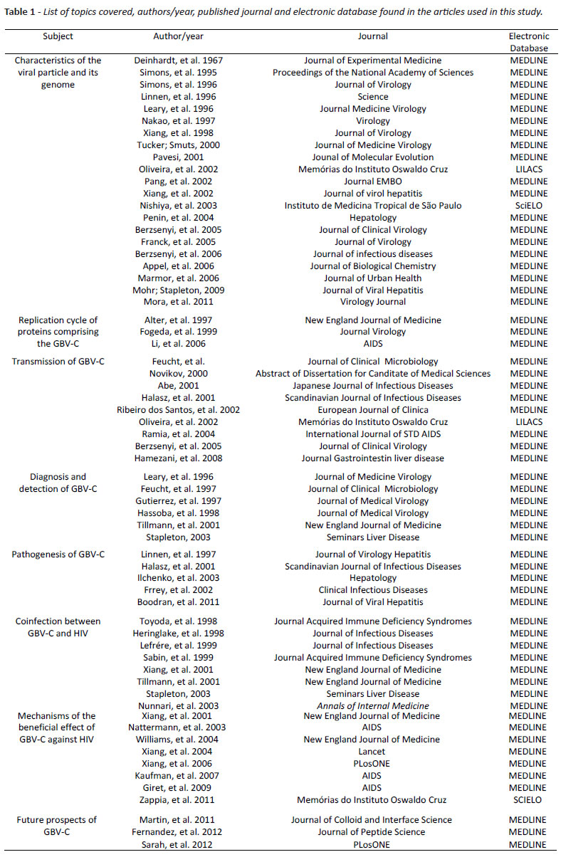
With respect to these 21 studies, two addressed the origin and evolution of GBV-C by phylogenetic analysis of their genotypes, two approached different genotypes of GBV-C in the world and two the different genotypes in Brazil. In the study of the replication cycle of proteins comprising the GBV-C, we use three studies, one of which refers to cell tropism of GBV-C and the other two of the possible replication cycle of the viral agent. The transmission of GBV-C was reviewed in nine articles, which approached parenteral, sexual and maternal and child routes. With respect to forms in the detection and diagnosis of GBV-C we used six studies reffering molecular and serological diagnosis. Regarding the pathogenesis, five studies were cited, where we study the behavior of GBV-C and the signs and symptoms in infected individuals. Co-infection between HIV and GBV-C was investigated in eight studies, which approached the beneficial effects of coinfection in different populations. The mechanisms used by GBV-C to exert a protective effect in HIV-infected individuals were studied in eight articles of our research, which suggest different mechanisms of interaction between GBV-C and HIV. The future prospects of the viral agent in the scientific community was found in three in vitro studies which test different proteins of GBV-C virus genome facing the ability to inhibit HIV replication. Table 1 illustrates among each subject studied, the authors used, year, journal published and electronic database where articles were found.
DISCUSSION Characteristics of the viral particle and its genome
Based on sequence and genome organization the GBV-C is classified as a member of the Flaviviridae family, which has three known genera: Flavivirus, Pestivirus and Hepacivirus(8). As illustrated in Figure 1, it is not currently assigned to the GBV-C none of the known genera, but it has been proposed that it be assigned to the Hepacivirus genus(8).
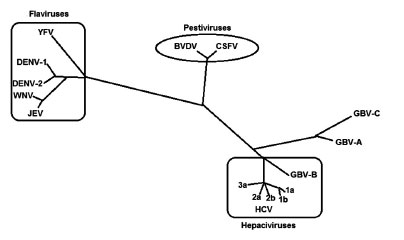 Figura 1 - Phylogenetic relationship of the family Flaviviridae.
GBV-C is a RNA viruses represented by a single stranded RNA with positive polarity(9). Its genome has 9.4 Kb and is organized similarly to HCV as shown in Figure 2(10,11). The different isolated viral genomes of GBV-C have between 9103 and 9392 nucleotides and contains one open reading frame (ORF) that encodes a large polyprotein precursor from 2873 to 2910 amino acids(10,11). The open reading frame is located between untranslated regions (UTR) at the 5' and 3' ends of the viral genome(6).
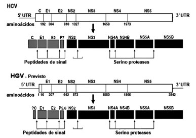 Figura 2 - Genomic organization and proteolytic processing of
the HCV and GBV-C(8) virus. With adaptations.
The 5' UTR of GBV-C is highly conserved and is higher than that of HCV, presenting an internal site of the ribosome entry (IRES, internal ribosomal entry site), which is responsible for starting the translation of viral RNA(12). The IRES are essential for translation of the viral genome and allow the direct connection of the 40s subunit of the ribosome, requiring no pre-boot factor(12). The IRES activity is lower in GBV-C than in HCV(10).
The translated viral polyprotein is cleaved by a combination of proteases from the host cell and viral pro-teases to then form the viral structural proteins and viral enzymes(13). The structural proteins of GBV-C are encoded in the n-terminal region of the ORF(13). The structural proteins is a capsid derived whose coding regions has not yet been completely identified(14), two envelope glycoproteins, E1 and E2 forming a heterodimer and are located on the surface of the viral particle, and an ion channel (p5.6)14), as shown in Figure 2(8).
The C-terminal portion of ORF of GBV-C encodes nonstructural proteins which are NS2 , NS3 , NS4A, NS4B, NS5A and NS5B(15), as shown in Figure 2(8). Although not completely characterized experimentally, it is believed that their functions are analogous to HCV proteins(15). Thus, after cleavage by cellular peptidases, NS2 is first protease activated(16). Its C-terminal portion contains a protease domain, which together with the n-terminal domain of NS3 catalyze the cleavage between NS2 and NS3(16). It is believed that in the GBV-C the NS2 functions, such as: inhibition of apoptosis, modulation of gene expression and NS5A phosphorylation(16). NS3 is versatile and has serine protease activity, RNA helicase and nucleotidase (nTPase)(17). Its helicase domain is related to the initiation of RNA synthesis by dissociation of nascent RNA tapes and their molds(17). NS4A is a cofactor of the serine protease and is responsible for NS3/NS4A cleavage of all subsequent proteins(12, 13, 14, 18, 19, 20). The NS5A and NS4B are poorly characterized, but it is known that NS4B protein recruits other components of the replicase to the site of viral replication and that NS5A interacts with the innate immune system(12, 13, 14, 18, 19, 20). The NS5B is the RNA-dependent RNA polymerase, which is essential for RNA replication machinery(12, 13, 14, 18, 19, 20).
Using samples from around the world, the phylogenetic analysis of the genomic sequences of subtype GBV-C revealed the existence of six genotypes(1, 2, 3, 4, 5, 6), directly related to the geographic region of origin, suggesting that GBV-C is an ancient virus that was disseminated to the migration of the population over the years(4,21).
The genetic diversity of GBV-C varies according to the different regions where the virus was detected; genotype 1 predominates mainly in West Africa, genotype 2 in Europe and USA, genotype 3 in Asia, genotype 4 in the Southeast Asian, genotype 5 in South Africa and genotype 6 in Indonesia(22).
In a study of blood donors in Colombia, we assessed 408 individuals, with a prevalence of 5.06% of GBV-C infection(23). Of these, 41.6% had a genotype 2a, 33.3% the genotype 1, 16.6% the genotype 3 and genotype 2b 8.3%(23).
In Brazil, few studies have been performed(24, 25).Most of them noted the predominance of genotypes 1 and 2 with 8.3 to 17.6 % and from 82.4 to 91.7% of prevalence respectively(24, 25).
GBV-C is the human virus most similar to HCV(6, 15). Through genomic sequence analysis we found a homology of about 30% of their amino acids(6, 15). A gene region that has the highest homology is NS3, or the coding region of the viral helicase(6, 15). The gene portions encoding the viral envelope proteins E1 and E2 have the highest differences between these viruses(6, 15).
Replication cycle proteins comprising the GBV-C
The replication cycle of the proteins that composes the GBV-C has not been clarified, it is believed that replication occurs in the cytoplasm, the same way it occurs the flavivirus replication of single-stranded RNA with positive polarity by the synthesis of a RNA molecule of negative polarity, which serves as a sample for the production of new positive tapes(26).
The GBV-C, despite being called hepatitis G virus, has little or no tropism for hepatocytes and hepatic replication is little described(27). These findings were demonstrated by alter et al., in 1997, by inoculating the virus in primates chimpanzees and failed to show signs of liver disease(27).
The GBV-C has tropism for lymphocytes and their replication occurs early in mononuclear cells of peripheral blood as T CD4+ lymphocytes, B lymphocytes, bone marrow and spleen(28).
Transmission of GBV-C
Infection with GBV-C is common throughout the world, and can be transmitted by parenteral, sexual and maternal and child routes(5, 29, 30).
The parenteral route is the most important, mainly through blood transfusion and blood products(5). The GBVC has high rates of prevalence in injecting drug users, hemophiliacs, hemodialysis and transplant patients(5).
Indirect evidence of parenteral transmission of GBV-C are higher frequencies detected of this virus in groups at high risk of infection for hepatitis B and C who have similar ways of transmission(31).
In a study performed by Berzsenyi et al., in 2005 were assessed individuals considered healthy, with no risk factors for sexually transmitted diseases and normal transaminases rates(6). The study found 1.9% of patients positive for GBV-C(6). Of these individuals, 6.8% were on hemodialysis, 18.2% were HIV positive, 24.4% were HCV positive, 28.8% were intravenous drug users, 21.1% had received a blood transfusion and 35.2% were hemophiliacs(6). These findings indicate that the prevalence of GBVC varies according to the population studied(6).
Few studies on the transmission of GBV-C were performed in Brazil(25). In the state of Goiás, a study was performed in order to investigate the prevalence of GBV-C in 241 blood donors(25). Of these, 17 (7.1%) were positive for GBV-C(25).
Sexual transmission of GBV-C is of great importance. In research performed by Hamezani, et al., in 2008 in iran, were assessed the frequency of GBV-C in 82 patients infected with HIV(32). The researchers found 10.97% prevalence of coinfection GBV-C/HIV and of these co-infected subjects, 13.7% had only risk factors for transmission and 6.7% parenteral risk factors only for sexual transmission(32). This study suggests that sexual transmission is very efficient in individuals without risk factors for parenteral transmission(32).
A study performed in Brazil with 1,039 individuals in the city of São Paulo showed a greater prevalence of GBV-C and sexually active individuals who perform risky sexual practices, such as men who have sex with men(33).
The maternal-infant transmission of GBV-C has also been documented, a survey that examined African children with mothers positive for GBV-C found rates of 15% of children infected by the same virus(5). Halasz et al., in 2001 found 89% of vertical transmission, suggesting however that more research should be performed to verify the persistence of GBV-C and spontaneous healing in children infected by this route of transmission(34).
Diagnosis and detection of GBV-C
Most asymptomatic individuals infected with GBV-C clear the virus after development of antibodies(35). These antibodies are produced against the viral envelope glycoprotein E2 (anti-E2)(35). The encountering of these antibodies is indicative factor that the individual has been exposed to the virus(29, 36). Immunoenzymatic techniques (eLiSa) are used to detect the presence of anti-E2, indicating bleaching and immunization against the viral reinfection(29, 36). Due to be an anti-E2 neutralizing antibody, if present, the subject is unlikely to present viremia, which occurs only in 1% of the cases, suggesting that anti-E2 is associated with previous infection(37).
The molecular diagnostics for detecting exposure to GBV-C is also of great importance. Molecular biology tests such as RT-PCR (reverse transcription polymerase chain reaction) lead to the detection of viral RNA in serum or plasma from patient suspected(15, 37). The technique of viral nucleic acid amplification by RT-PCR, nested PCR or real time - PCR use mostly the method of 5' UTR, NS3 and nS5 because they are the most conserved regions of the genome do GBV-C(15, 37). The finding of viral RNA is indicative of virema or active infection(15, 37). According the mentioned above, it would be of great importance that studies aiming at estimating the exposure GBV-C would use both methods for detection of RNA GBV-C and antiE2(15, 37). A study of GBV-C virus performed by Hassoba et al., in 1998, studied if a group of positive anti-E2 patients in need of liver transplant, continue with this antibody after transplantation, and if the anti-E2-positive patients after transplantation were protected against GBV-C against re-infection(38). The researchers concluded that although liver transplantation cause immunosuppression, the antiE2 remained being detected in positive individuals earlier. Further, they found that the presence of anti-E2 appear to prevent reinfection(38).
Pathogenesis
The mechanism responsible for the development of hepatitis induced by GBV-C is not clear today. Although this virus have been discovered in a patient with hepatitis and existing case reports of acute and chronic hepatitis G, the hepatotropism remains controversial(39).
Halasz, et al., in 2001, through a histological research performed on the liver of patients infected with this viral agent believe that there is no association between the presence of GBV-C and changes in hepatic cells(34). The same results were found by Boodram, et al., in 2011(40).
Even facing these conflicts in the literature regarding the pathogenicity of hepatitis G virus, most authors believe that GBV-C is probably not virulent or a virus associated with severe liver disease; it does not impair the liver disease already installed and also that the liver is not its primary site of infection(41).
Therefore, studies suggest that GBV-C leads to development of liver injury in infected individuals are contradictory, and it was not possible to prove that this association exists(42). For this reason, Linnen et al., in 1997 believed that through the literature data the nomenclature "hepatitis G (GBV-C)" can be replaced only by GB-C virus(42).
GBV-C co-infection with HIV
Studies available show that co-infection of GBV-C and HIV brings favorable prognosis for HIV-infected patients, with the progression of HIV to AIDS more slowly with increased rates of T CD4+ cells providing greater survival at HIV-infected individuals(2,3,7).
HIV is transmitted by sexual, parenteral and maternal and child routes, and such routes also disseminate other agents, including GBV-C, which explains the high rate of GBV-C infection in HIV-positive patients(43).
Today, GBV-C coinfection with HIV is considered frequent, since as mentioned earlier these two viruses have efficient transmission by sexual and parenteral routes highlighting the importance of studies to assess this association(37).
The first study relating GBV-C and HIV was performed by Toyoda et al. 1998 in Japan, attracting the interest of the scientific community on this coinfection(2). The survey was performed with 41 hemophiliacs infected with HIV by blood transfusion(2). The researchers besides having found a 27% prevalence of GBV-C coinfection and HIV, suggested the beneficial effect of GBV-C in HIV-positive patients, since these patients showed a decrease in HIV viral load(2). Despite this finding, the researchers failed to demonstrate that the GBV-C would lead to a decrease in time to progression to AIDS, but opened up a range for further studies relating the GBV-C and HIV(2).
Also in the same period, Heringlake, et al. 1998 performed a survey of 197 individuals from Germany, positive for HIV, which sought to verify the influence of the GBV-C in HIV patients group(3). The infection to GBV-C was found in 16.8% of HIV-infected individuals, whereas the prevalence of anti-E2 was 56.8%(3). The researchers also found that HIV-positive patients with active infection of GBV-C (16.8%) had a count of T CD4+ cells higher than the positive anti-E2 and negative for GBV-C(3).
Studies performed by Sabin, et al.,in 1999 assessed the mortality of 94 British hemophiliacs co-infected with GBV-C and HIV (44). The researchers found no significant difference on the influence GBV-C on the mortality of patients infected with HIV(44). One should take into consideration that this study did not differentiate the patients with active infection of GBV- C from those who showed anti-E2, and all patients were considered as exposed to the GBV-C virus(44).
In contrast to the team of Sabin, in the same year, Lefrére, et al., in 1999, confirmed the results found by Toyoda and Heringlake in 1998 in a survey performed in approximately five years, with 97 French-infected HIV patients(43). The study revealed that the group infected with GBV-C had larger T CD4+ counting, with a less progression to AIDS emphasizing that the beneficial effect of GBV-C in HIV-positive population(43). Furthermore, it was observed that among the patients with active infection of GBV-C, only 39% required to start antiretroviral therapy, whereas 68% of patients infected only with HIV needed antiretroviral drugs(43).
Following the same line of research, Tillmann, et al., in 2001 performed a study with 197 patients with HIV, using antiretroviral therapy (arT)(36). The researchers sought to elucidate the relationship between long-term GBVC and patients infected with HIV-AIDS(36).
The findings showed that patients positive for RNA-GBV-C had a lower HIV viral load with a slower advance to the disease than patients mono-infected with HIV(36). Furthermore, it was observed that individuals using arT co-infected with GBV-C also showed beneficial effects on their survival, an increase in the T CD4+ cells counting(36). With these findings researchers have confirmed that the relationship between the GBV-C virus and HIV is associated with reduced mortality in HIV-infected patients, even after the introduction of HaarT, suggesting that the presence of GBV-C inhibits HIV replication(36).
Nunnari, et al. 2003 confirmed the study of Tillmann et al. 2001 through findings that GBV-C would lead to increased patient survival, either before or after initiation of HaarT, with improvement in response of this therapy(36, 45). This study reinforced the findings regarding the beneficial effect on HIV patients co-infected with GBV-C(45).
Mechanisms of the beneficial effect of GBV-C against HIV
Although the studies are controversial, there is strong evidence that GBV-C somehow may inhibit the replication of HIV, where there are several hypotheses about the possible mechanisms by which this GBV-C exerts a protective effect, but these mechanisms are not completely elucidated(46).
Williams et al. 2004 studied 271 patients who were assessed from seroconversion for a period of 11 years through the analysis of infection and the presence of anti-E2(46). It was found that GBV-C infection in HIV patients leads to increased survival in subjects with higher period of GBV-C infection and insofar presented clearing of GBVC, the patients had a worsening of the HIV infection(46). In this study the researchers suggested that these beneficial results are dependent on the time of HIV infection, or that is, the longer the duration of HIV infection the more the GBV-C will exert a protective effect, the fact that the opposite results clarify the protective effect of GBV-C in other studies(46). Possible mechanisms of the protective effect of GBV-C against HIV infection are described below.
Inhibition of HIV replication
One of the hypotheses trying to explain the way in which the GBV-C minimizes the progression of HIV would be a direct inhibitory action of GBV-C in HIV replication, due to the fact that both viruses replicate in human peripheral blood mononuclear cells including on T CD4+, T CD8+ lymphocytes and B lymphocytes(7). This coinfection could affect the initial stages of the cycle of HIV binding and fusion to the target cell, reverse transcription, and then integrating into the genome of the host cell, production of the provirus, among other, affecting HIV replication without increasing cellular toxicity(7).
Induction of a cytokine profile favoring HIV infection
Another hypothesis is that GBV-C/HIV coinfection would stimulate a profile of positive immune response against HIV infection, inducing an increase in Th1 response through the production of IL-2, IL-12 and IFN gamma, by mononuclear cells stimulating cellular immune response(47).
Decreased expression of chemokine receptors
We also found that the mechanism of GBV-C in HIV progression has relation with the CCR5 and CXCR4 chemokine receptors, which are very important for the occurrence and progression of HIV transmission(48). Some strains of HIV utilize the CCR5 receptor for entry into target cell, while others use the CXCR4 co-receptor(48). In a study published by Xiang et al., in 2004, an in vitro study was performed where researchers inoculated GBV-C and HIV viruses in mononuclear cells(48).
The presence of GBV-C provides secretion of cytokines such as ranTeS, MIP-1α, MiP-1β, natural ligands of CCR5 and SDF-1, thus reducing the expression of CD4, CCR5 and CXCR4 used by HIV for cell entry, and also found that co-infection led to an 23% reduction in HIV replication(48).
In a study by nattermann, et al., in 2003, they assessed the expression of a cell receptor utilized by HIV, CCR5, in lymphocytes from HIV negative and positive patients with viremic and non viremic patients for GBV-C(49). The findings showed that GBV-C led to an increased secretion of cytokines and decreased expression of CCR5, which would explain why GBV-C lessen the progression of HIV(49).
In a study published by Giret, et al., in 2009, they assessed the effect of GBV-C infection in the activation of T cells in 48 patients with recent HIV infection(50). The findings confirmed the positive effects between the viral load of HIV-1 and rates of T CD8+ and CD38(50). It was also demonstrated that the GBV-C virus infection resulted in a decreased expression of CD38 on T CD4+ and T CD8+ and CCR5 cells on T CD8+ cells(50). The results were independent of the viral load of HIV-1, the count of T CD4+ and T CD8+ cells(50). The authors stated further that the GBV-C virus infection is associated with less activation of T cells, and this can be a key mechanism for protecting the HIVinfected patient, thereby reducing disease progression(50).
In contrast to the previous study, Zaapia, et al., in 2011, performed a study in a hospital in the city of São Paulo, which assessed the effect of GBV-C on the immune response to HIV(51). They assessed 159 individuals with chronic HIV infection, and 65 (40.8%) of these had markers of GBV-C infection(51). Despite the high prevalence, the presence of GBV-C had no effect on HIV replication in T CD4+ and CD8+ cell counting(51). The immune response by IFN-g, IL-2 and CD38 did not differ between the groups co-infected with GBV-C/HIV and only HIV-positive(51). The authors did not exclude the protective effect of GBV-C in early HIV infection, but they did not find this beneficial effect in patients coinfected with HIV in chronic phase(51).
It was observed that the E2 protein of GBV-C binds CD81 cells, promoting the release of ranTeS, with subsequent internalization of CCR5 receptor of mononuclear cells by decreasing their expression on the surface of lymphocytes, which may block the HIV entry in the host cell(52).
Different genotypes exert different effects
Xiang in 2006 found that the non-structural protein NS5A of GBV-C has inhibitory action on the CXCR4 co-receptor by increasing its SDF-1 ligand, which reduces the entry of HIV into the host cell(47). Moreover, the authors found that when after the expression of NS5A of GBV-C, only genotypes 1, 2, 3 and 5 were able to inhibit the replication of HIV, suggesting that different genotypes may exert different effects on the development of HIV infection(47).
Future perspectives on the GBV-C
Recently, Martin et al., in 2011, performed an in vitro study to identify proteins in GBV-C which performs the function of inhibiting the activity of the gp41 surface protein of HIV, being this the fusion peptide of HIV into the target cell (53). Were synthesized and studied 58 GBV-C peptides corresponding proteins E1 envelope (53). Of these synthetic peptides, five showed a reduction in fusion activity of gp41 to a synthesized liposomal model membranes similar to membrane of the target cell(53). The authors believe that these synthetic peptides may contribute to the development new therapeutic agents for the treatment of AIDS(53).
Another study with synthetic peptides derived from GBV-C was performed by Fernandez et al., in 2012(54). The authors assessed the activity of a synthetically modified peptide derived from the E2 region of GBV-C, compared to gp41 from HIV(54). The study revealed that the modifications made to the peptide selected did not inhibit fusion of the 41 gp to the target cells(54). Studies with synthetic peptides are recent and despite being controversial, open a range for the scientific community to seek new alternatives for HIV treatment, and a better understanding of the GBV-C viral agent(53, 54).
Within this context, Sarah, et al., in 2012(55)performed an in vitro investigation of genome sequence of GBVC encoding a serine protease (NS3) assessing whether this protease somehow affects the cellular environment of lymphocyte by impairing the replication of HIV. Researchers found that when the NS3 protease of the GBV-C genome was expressed in a strain of human T CD4+ lymphocytes, the HIV replication in these lymphocytes significantly decreased, and this reduction in replication was dependent from the NS3 dose tested. Moreover, the presence of NS3 of GBV-C on lymphocytes tested did not inhibit the expression of CD4 e CXCR4 lymphocyte receptors that are used by HIV entry to the host cell.
CONCLUSION
Therefore, even if the mechanisms leading to protective effect of GBV-C are controversial in the literature, scientific evidence lead us to believe that the GBV-C brings a improvement in the prognosis of patients infected with HIV. The hepatitis G virus appears to be distributed throughout the world population and it is believed that the prevalence is greater in individuals exposed to blood and blood products.
The GBV-C does not have great clinical impact in infected patients and does not cause severe liver disease, which leads to a detachment in detection of the viral agent and its antibodies in routine examinations. For this reason, in addition to the few specific studies of prevalence, we suppose that this virus is under-diagnosed and its prevalence underestimated in most regions of Brazil and the world.
Studies indicate that coinfection between GBV-C and HIV is a favorable prognostic factor for HIV-positive patients, deserving further investigation in the area, both approaching the prevalence of GBV-C co-infection with HIV and the possible mechanisms of viral interference. We know several regions in our country of high HIV prevalence, where genetic variation of the virus is the target of interest to researchers. Through these studies we may have more information on GBV-C and its effects on HIV-positive patients. Current research of GBV-C that perform in vitro test for the capacity of different proteins forming part of its genome in inhibiting the replication of HIV is something promising for the population. Given that the resistance to antiviral drugs is a problem faced for many HIV-positive patients, the development of antiviral drugs to contribute with new treatment strategies for HIV is needed.
REFERENCES
1. Deinhardt F, Homes AW, Capps RB, et al. Studies on the transmission of human viral hepatitis to marmoset monkeys. Transmission of disease, serial passages and description of liver lesions. Journal of experimental Medicine. 1967, 125:673-88.
2. Toyoda H, Fukuda Y, Hayakawa T, et al. Effect of GB virus C/hepatitis G virus co-infection on the course of HIV infection in hemophilia patients in Japan. Journal acquired immune Deficiency Syndromes. 1998, 17:209-13.
3. Heringlake S, Ockenga J, Tillmann HL, et al. GB virus C/hepatitis G virus infection: a favorable prognostic factor in human immunodeficiency virus-infected patients? Journal of infectious Diseases. 1998, 177:1723-6.
4. Pavesi A. Origin and evolution of GBV-C/Hepatitis G virus and reationships with ancient human migrations. Jounal of Molecular evolution. 2001, 53:104-13.
5. Abe K. GB virus C/hepatitis G virus. Japanese Journal of infectious Diseases. 2001, 54:55-63.
6. Berzsenyi MD, Bowden DS, Roberts SK. GB virus C: insights into coinfection. Journal of Clinical Virology. 2005, 33:257-66.
7. Xiang J, Wünschmann S, Diekema DJ, et al. Effect of co-infection with GB virus C (hepatitis G virus) on survival among patients with HIV infection. new england Journal of Medicine. 2001, 345:707-14.
8. Mohr EL, Stapleton JT. GB virus type C interactions with HIV: the role of envelope glycoproteins. Journal of viral hepatitis. 2009,16:757-68.
9. Nakao H, Okamoto H, Fukuda M, et al. Mutation rate of GB virus C/hepatitis G virus over the entire genome and in subgenomic regions. Virology. 1997, 233:43-50.
10. Simons JN, Desai SM, Schultz DE, et al. Translation initiation in GB viruses a and C: evidence for internal ribosome entry and implications for genomic organization. Journal of Virology. 1996, 70:6126-35.
11. Linnen J, Wages JJ, Zhang-keck ZY, et al. Molecular cloning and disease association of hepatitis C virus: a transfusion-transmissible agent. Science. 1996, 271:505-8.
12. Appel N, Schaller T, Penin F, et al. From structure to function: new insights into hepatitis C virus RNA replication. Journal Biological Chemistry. 2006, 281:9833-36.
13. Penin F, Dubuisson J, Rey FA, et al. Structural biology of hepatitis C virus. Hepatology. 2004, 39:5-19.
14. Xiang J, Klinzman D, Mclinden J, et al. Characterization of hepatitis G virus (GB-C virus) particles: evidence for a nucleocapsid and expression of sequences upstream of the E1 protein. Journal of Virology. 1998, 72:2738-44.
15. Leary TP, Muerhoff AS, Simons JN. Sequence and genomic organization of GBV-C: a novel member of the Flaviviridae associated with human non a-e hepatitis. Journal Medicine Virology. 1996, 48:60-7.
16. Franck N, Leseyec J, Guguen GC, et al. Hepatitis C virus NS2 protein is phosphorylated by the protein kinase CK2 and targeted for degradation to the proteasome. Journal of Virology. 2005, 79:2700-8.
17. Pang PS, Jankowsky E, Planet PJ, et al. The hepatitis C viral NS3 protein is a processive Dna helicase with cofactor enhanced RNA unwinding. Journal EMBO. 2002, 21:1168-76.
18. Marmor M, Hertzmark K, Thomas SM, et al. Resistance to HIV infection. Jounal of Urban Health. 2006, 83:5-17.
19. Xiang J, Daniels KJ, Soll DR, et al. Visualization and characterization of GB virus C particles: evidence for a nucleocapsid. Journal of virol hepatitis. 2002, 6:16-22.
20. Simons JN, Pilot-matias TJ, Leary TP, et al. Identification of two flavivirus-like genomes in the GB hepatitis agent. Proceedings of the National academy of Sciences. 1995, 92:3401-05.
21. Berzsenyi MD, Roberts SK. What is the role do GB virus C infection in hepatitis C virus/HIV coinfection? Journal of infectious diseases. 2006, 194:407-9.
22. Tucker TJ, Smuts HE. GBV-C/GBV-C genotypes: proposed nomenclature for genotypes 1-5. Journal of Medicine Virology. 2000, 62:82-3.
23. Mora AVM, Botelho L, Nishiya A, et al. Frequency and genotypic distribuition of GB vírus C (GBV-C) among Colombian population with hepatitis B (HBV) or hepatitis C (HCV) infection. Virology Journal. 2011, 8:345.
24. Nishiya AS, Ribeiro-dos-Santos G, Bassit L, et al. Genotype distribution of the GB virus C in citizens of São Paulo City Brazil. Instituto de Medicina Tropical de São Paulo. 2003, 45:213-6.
25. Oliveira LA, Martins RM, Carneiro MA, et al. Prevalence and genotypes of GB virus C/hepatitis G virus among blood donors in central Brazil. Memórias do Instituto Oswaldo Cruz. 2002, 97:953-7.
26. Fogeda M, Navas S, Martin J, et al. In vitro infection of human peripheral blood mononuclear cells by GB virus C/hepatitis G virus. Journal virology. 1999, 73:4052-61.
27. Alter MJ, Gallagher M, Morris TT, et al. Acute non-A-E Hepatitis in the United States and the role of Hepatitis G virus infection. New england Journal of Medicine. 1997, 336:741-6.
28. Li C, Collini P, Danso K, et al. GB virus C and HIV-1 RNA load in single virus and co-infected West African individuals. AIDS. 2006, 20:379-86.
29. Feucht HH, Zollner B, Polywka S, et al. Prevalence of hepatitis G viremia among healthy subjetcs, individuals with liver disease, and persons at risk of parenteral transmission. Journal of Clinical Microbiology. 1997, 35:767-8.
30. Ramia S, Mokhbat J, Sibai A, et al. Exposure rates to hepatitis C and G virus infections among HIV-infected patients: evidence of efficient transmission of GBV-C by the sexual route. International Journal of STD AIDS. 2004, 15:463-6.
31. Novikov DV. Molecular Biological characteristics of HCV. Abstract of dissertation for Canditate of Medical Sciences. 2000, 1-22.
32. Hamezani A, Mohraz M, Vahabpour R, et al. Frequency of hepatitis G virus infection among HIV positive subjects with parenteral and sexual exposure. Journal Gastrointestin liver disease. 2008,17:267-72.
33. Ribeiro-dos-Santos G, Nishiya AS, Nascimento CM, et al. Prevalence of GB virus C (hepatitis G virus) and risk factors for infection in São Paulo, Brasil. European Journal of Clinical. 2002, 21:438-43.
34. Halasz R, Weilanda O, Sällberg M. GBV virus C/hepatitis G virus. Scandinavian Journal of infectious Diseases. 2001, 33:572-80.
35. Gutierrez RA, Dawson GJ, Knigge MF, et al. Seroprevalence of GB virus C and persistence of RNA and antibody. Journal of Medical Virology. 1997, 53:167-73.
36. Tillmann HL, Heiken H, Knapik-botor A, et al. Infection with GB virus C and reducet mortality among HIV-co-infected patients. New England Journal of Medicine. 2001, 345:715-24.
37. Stapleton JT. GB virus type C/hepatitis G virus. Sem Liver Dis. 2003, 23:137-48.
38. Hassoba HM, Pessoa MG, Terrault NA, et al. Antienvelope antibodies are protective against GBV-C reinfection: evidence from the liver transplant model. Journal of Medical Virology. 1998, 56:253-8.
39. Ilchenko LY, Sharafanova TI, Shepeleva SD, et al. Antibodies to Hepatitis G virus in patients with chronic liver diseases. Hepatology. 2003, 5:4-6.
40. Boodram B, Hershow CR, Klinzman D, et al. GB virus C infection among young, HIV-negative injection drug user with and without hepatitis C virus infection. Journal of viral hepatitis. 2011, 18:153-9.
41. Frey SE, Himan SM, Sokol-anderson M, et al. Evidence for probable sexual transmission of the hepatitis G virus. Clinical infectious Diseases. 2002, 34:1033-8.
42. Linnen JM, Fung K, Fry KE, et al. Sequence variation and phylogenetic analysis of 5 terminus of hepatitis G virus. Journal of Virology Hepatitis. 1997, 48:2771-7.
43. Lefrére JJ, Roudot-thotaval F, Morand-joubert L, et al. Carriage of GB virus C/hepatitis G virus rna is associated with a slower immunologic, virologic and clinical progression of human immunodeficiency virus disease in co-infected persons. Journal of infectious Diseases. 1999, 179:783-9.
44. Sabin CA, Devereux H, Klinson Z, et al. Effect of co-infection with hepatitis G virus on HIV disease progression in hemophilic men. Journal acquired immune Deficiency Syndromes. 1999, 19:346-7.
45. Nunnari G, Nogro L, Palermo F, et al. Slower progression of HIV-1 infection in persons with GB virus C co-infection correlates with an intact T-helper 1 cytokine profile. Annals of internal Medicine. 2003, 139:26-30.
46. Williams CF, Klinzman D, Yamashita TE, et al. Persistent GB virus C infection an survival in HIV infected men. New england Journal of Medicine. 2004, 350:981-90.
47. Xiang J, Mclinden JH, Chang Q, et al. Characterization of a peptide domain within the GB virus C NS5A phosphoprotein that inhibits HIV replication. PLosone. 2006, 3:1-10.
48. Xiang J, George SL, Wünschmann S., et al. Inhibition of HIV-1 replication by GB virus C infection through increases in ranTeS, MIP-1α, MIP-1β, and SDF-1. Lancet. 2004, 363:2040-6.
49. Nattermann J, Nischalke HD, Kupfer B, et al. Regulation of CC chemokine receptor 5 in hepatitis G virus infection. AIDS. 2003, 17:1457-62.
50. Giret MTM, Silva TM, Sauer MM, et al. GB virus type C infection modulates T-cell activation independently of HIV-1 viral load. AIDS. 2009, 23:2277-87.
51. Zappia BLG, Barbosa JL, Brunialti CKM, et al. Influence of GB vírus C on IFN-γ and IL-2 production and CD38 expression in T lymphocytes from chronically HIV-infected-HCV-co-infected patients. Memórias do Instituto Oswaldo Cruz. 2011, 106:662-9.
52. Kaufman TM, Mclindem JH, Xiang J, et al. The GBV-C envelope glycoprotein E2 does not interact specifically with CD81, AIDS. 2007, 21:1045-8.
53. Martin SJM, Hristova K, Pujol M, et al. Analysis of HIV-1 fusion peptide inhibition by synthetic peptides from E1 protein of GB virus C. Journal of Colloid and interface Science. 2011, 360:124-31.
54. Fernandez L, Chan CW, Egido M, et al. Synthetic peptides derived from an N-terminal domain of the E2 protein of GB virus C in the study of GBV-C/HIV-1 co-infection. Journal of Peptide Science. 2012, 18:326-35.
55. George LS, Varmaz D, Tavis EJ, et al. The GB Virus C (GBV-C) NS3 Serine Protease inhibits HIV-1 replication in a CD4+ T Lymphocyte Cell Line without Decreasing HIV receptor expression. Plosone. 2012, 7:1-8.
1. Especialista (Bioquímica e Mestranda) 2. Doutora (Bióloga e Doutora) 3. Mestre (Bióloga e Doutoranda) 4. Mestre (Bioquímica e Doutoranda) 5. Doutora (Doutora e Professora adjunta da Universidade Federal do Rio Grande)
Campus Saúde Rua: Visconde de Paranaguá 102 Centro Rio Grande/RS CEP único: 96203-900 Telefone para contato/fax: (53)3233.8800 E-mail:luisadias_sixcp@hotmail.com
Trabalho de revisão desenvolvido no curso de especialização em agentes infecto-parasitários de interesse humano no Programa de Pós-Graduação em Ciências da Saúde da Universidade Federal do Rio Grande (FURG)