INTRODUCTION
Dental occlusion is considered normal when the upper and lower teeth are aligned in their bony bases in the center of the alveolar ridge and in relative class I1. Thus, the upper dental arch needs to be proportionately greater than the lower arch, making the palatal cusps of the premolars and molars fit properly to occlusal pits of the premolars and mandibular first molar1. When there is any deviation from a morphological characteristics of normal occlusion and manifestation in the senses of space, including the transverse dimension, it is called malocclusion. The result of atresia of the upper dental arch in front of the inferior arch of normal dimensions is the posterior crossbite2.
The diagnosis of posterior cross bite is very easy, since the normal reference morphology is near, at the lower arch. Thus, we can define the posterior crossbite as the upper arch atresia without compensation of the lower arch. However, the posterior crossbite is not the only indicator of maxillary atresia. The maxillary atresia may be present in the case of simultaneous atresia of the lower dental arch. This implies the absence of posterior cross bite. Thus, the cross-sectional diagnosis should be based on inter- and intra-arch relationship2,3.
For the treatment of maxillary atresia, rapid Maxillary expansion (RME) was described and originally performed by angell (1860)4 and later by Hass (1961)5 in the United States, and it was advocated that the ideal age range for the RME through orthodontic- orthopedic apparatus corresponds to the young patients, with maximum age ranging from 14 years for women and 16 for men, in which the median palatine sutures have not completed the ossifying process6.
In individuals who have reached skeletal maturity, the surgical separation is indicated in order to provide separation of the median palatine suture with consequent maxillary expansion and the decrease in ortodontic gradient effects7. This is known as Surgically assisted rapid Maxillary expansion (SARME) in which the use of a conventional expander is indispensable, and it may be tooth-supported or tooth-mucous-supported with the activator screw should be appropriate to the amount of expansion required. It can be performed under general anesthesia or local anesthesia, and this procedure is aimed at separating the sutures to prevent palate disjunction, through the techniques proposed by epker and Wolford (1980)8in which an osteotomy of Le Fort I type is performed associated with a osteotomy with chisel and hammer of pterigomaxilares sutures (not performed in cases under local anesthesia) and median palatine sutures (Figure 1). Postoperatively, the patient promotes the daily activation of the expansion device, up to the over-expansion controlled by the orthodontist, so that after completion of the process of bone repair (6 months), the professional proceeds with necessary orthodontic mechanics2,6,10,11.
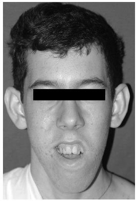 Figure 1 - Diagram showing the osteotomies performed during SARME.
OBJECTIVE
Given the dento-skeletal changes that maxillary atresia causes to patients, as well as the lack of technique and its indications, such issues justifies this study, by discussing them through a clinical case treated by Surgically assisted rapid Maxillary expansion (SARME).
METHODS AND RESULTS
Male patient, leucoderma, 19 years old, sought outpatient accompanied by his father, with referred by an orthodontist for evaluation of severe malocclusion. The patient had a medical history of arthrogryposis, a disorder characterized by multiple congenital rigid deformities of joints, which limits the locomotor development.
On extraoral examination, the patient had lip incompetence, facial pattern III, transverse deficiency and vertical maxillary excess, combined with horizontal mandibular excess (Figure 2).
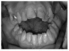 Figure 2 – Facial aspect in which the patient had lip incompetence, III facial pattern with horizontal maxillary deficiency, combined with horizontal mandibular excess.
In the intraoral examination, it was noted posterior and anterior crossbite, Class III and anterior open bite (Figure 3). In the analysis of models of inter-arch study, leading them from the occlusion clinically observed for Class I occlusion, we confirmed the absolute cross bite, with a severe maxillary atresia.
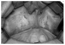 Figure 3 - In the intraoral examination, anterior and posterior cross bite , Class III and anterior open bite.
We proposed SARME under general anesthesia in a hospital for correction of maxillary atresia. First, the orthodontist installed the tooth-supported device (Hyrax) with screw of 13 mm long.
The surgery began with incision and mucoperiosteal detachment in the region of the upper gengivolabial groove (distal 16-26) (Figure 4). Then the billateral osteotomy from the nasal aperture to maxilla wall was performed using reciprocating saw (Striker®)5 mm above the apex of the teeth (Figure 5). Then, separation of the nasal septum (cartilage and vomer bone) using chisel with guide and hammer (Figure 6) was performed. At this time, using his index finger, the surgeon should palpate the boundary between the hard palate and soft palate, so that during the separation of the septum up to the posterior limit it can be noticed by the surgeon.
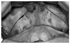 Figure 4 – Jaw access, followed by incision and mucoperiosteal detachment in the region of the upper gengivolabial groove.
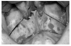 Figure 5 – Osteotomy using reciprocating saw (Striker®) from the nasal aperture to the jaw posterior wall bilaterally.
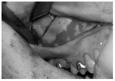 Figure 6 - Osteotomy of the nasal septum using chisel with guide and hammer.
The pterygomaxillary disjunction was performed bilaterally with curved chisel and hammer, blind, and the index finger of the opposite hand of the surgeon palpates pterygoid hamulus and maxillary tuberosity, in order to feel the separation of these structures when the disjunction of the pterygomaxillary suture occurs. During this osteotomy the chisel should remain at all times under the periosteum and be directed from lateral to medial and upper to bottom (Figure 7).
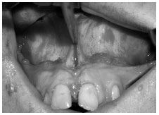 Figure 7 - Pterygomaxillary disjunction bilaterally with curved chisel and hammer.
At this time, the expanding device is activated (8-fold), which corresponds to 2 mm of expansion, with the intent of maintaining a tension in the palate. Then, a straight chisel was placed on the median palatine suture between the maxillary central incisors, and application of blows with the hammer. Clinically, when it occurs the separation of the jaws, one inter- spacing diastema is noticed, which is enhanced by prior activation of the expanding device (Figure 8).
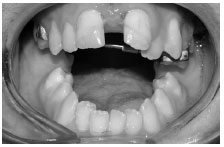 Figure 8 - Osteotomy of the median palatine sutures. The maxillary expansion with interincisor diastema is noted.
On the second day after surgery the patient was instructed on daily activations being 0.5 mm expansion per day until over-expansion is achieved. The screw was locked using acrylic resin for a period up to 6 months after installation of braces. Still, as treatment plan orthodontic mechanics for decompensation and alignment and leveling of the teeth will be performed, and later orthognathic surgery.
DISCUSSION
One of the most important aspects in the maxillary atresia is the correct diagnosis. The maxillary expansion is shown in transverse maxillary deficiencies, either absolute or relative, regardless of the stage of development of occlusion, from the deciduous dentition, since in the presence of posterior crossbite. When there is no posterior crossbite, transverse mechanics may start from the mixed dentition. However, there is a limit to the predictability of the outcome in relation to orthopedic patient age. From the clinical point of view, it is clear that the procedure of orthopedic maxillary expansion is fairly predictable until late adolescence. Thereafter, it becomes unpredictable and unlikely with increasing age. Given this circumstance, there is the option of eliminating the structural strength of the maxilla through osteotomies through the side walls and above the jaws (which break the zygomatic and canines buttress), median palatine sutures and pterigomaxillary suture, favoring the maxillary dysfunction even after adolescence2, 5, 10.
Therefore, in addition to clinical evaluation, analysis of dental casts is paramount. This is because it is necessary to determine whether the problem is dental or skeletal, and whether transversal deficiency is absolute or relative. The absolute deficiency is characterized by unilateral or bilateral crossbite after evaluation of plaster models with respect to Class I, often perceived in the evaluation of patients with retrognathia. In transversal relative disability, when the study models are placed in Class I, posterior crossbite is not observed, commonly seen in the assessment of patients with dentofacial deformity with Class III malocclusion. In the first situation ortho-surgical intervention to correct these deformities is required, and in the second no surgical treatment for correction of maxillary transverse dimension is indicated1, 10, 13. In this report, the patient was 19 years old, and corroborated with intraoral clinical examination and of plaster models, then we found the absolute crossbite, a fact that fully justified the indication for SARME.
There is a great debate in the literature regarding the performance of this surgical procedure under general or local anesthesia with or without sedation. Glassman, nahiogian, Medway (1984)14 in order to perform this procedure on an outpatient basis, described SARME without osteotomy of the nasal septum and pterygomaxillary suture, in order to provide greater comfort and safety to patients because it is a less invasive technique. However, controversies have arisen to determine which one would be less invasive procedure to be performed without affecting the amount of expansion achieved and the stability of such expansion over time, especially in severe jaw atresia.
In this context, the authors of this study are consistent in indicating conservative approaches where possible, to avoid subjecting the patient to surgery under general anesthesia, which is more costly since the operation performed in a hospital environment presents additional costs of hospitalization of the patient and anesthetist's fees. However, we also agree that RESAM through the osteotomy of all skeletal reinforcement, ensures greater stability in the correction of transverse maxillary deficiency, and further expansion in the molar region. This time, based on this philosophy, we indicate to the patient of this study the surgery in a hospital environment. The genetic defect presented by the patient (arthrogryposis), not incapacitated him to undergo an operation of such magnitude, since it does not alter the systemic functions, but only the locomotor autonomy.
Regarding the surgical technique, the SARME is a procedure that requires from surgeon specific maxillofacial training, in view of the possible complications. The main complications are vascular lesions during disjunction of pterygomaxillary suture due to disruption of the pterygoid venous plexus or maxillary artery branches2, 9. As well as the incorrect positioning of the chisel during osteotomy of the median palatine sutures, in cases on which the roots of the central incisors are very close it can result in fracture between the tooth root and the alveolar bone wall of central incisor15, 16. In this case there was no complications, and it is mandatory for any procedure the improved knowledge of the surgical technique by surgeons.
Based on the literature and presentation of clinical-surgical report, we conclude that the surgical technique provided good predictability in the correction of transverse deficiency, with minimal morbidity, the diagnosis by clinical evaluation and study models is essential for the indication of SARME; performing this surgery under local or general anesthesia will depend on the degree of maxillary atresia, the SARME before orthognathic Surgery will help orthodontic mechanics of decompensation, alignment and leveling of the teeth.
REFERENCES
1. Bratu DC, Bratu EA, Popa G, Luca M, Bălan R, Ogodescu A. Skeletal and dentoalveolar changes in the maxillary bone morphology using two-arm maxillary expander. Rom J Morphol embryol. 2012;53(1):35-40.
2. Lippold C, Stamm T, Meyer U, Végh a, Moiseenko T, Danesh G. Early treatment of posterior crossbite -a randomised clinical trial. Trials. 2013 Jan 22;14:20. doi: 10.1186/1745-6215-14-20.
3. Silva Filho O, Freitas SF, Cavassan AO. Prevalência de oclusão normal e maloclusão em escolares da cidade de Bauru (São Paulo): part. I relação sagital. Rev Odontol USP. 1990;4(2):130-7.
4. Angell EH. Treatment of irregularities of the permanent adult tooth. Menos invasivo a ser realizado sem prejudicar a quantidade Dent. Cosmos. 1860; 1: 540-4.
5. Hass AJ. Rapid expansion of the maxillary dental arch and nasal cavity by opening the midpalatal suture. Angle Orthod. 1961; 31: 73-90.
6. Rocha NS. et al., Discrepância transversal da maxila: tratamento ortodônticocirúrgico. Rev. Cir. Traumatol. BMF. 2005;5(2):55-60.
7. Bernardes LAA, Vieira PSR. Disjunção palatal cirurgicamente assistida: relato de caso. Rev. Clin. Ortod. Dent. Press. 2003;2(1):63-9.
8. Epker BN, Wolford LM. Dentofacial Deformities: surgical-orthodontic correction. St. Louis: Ed. Mosby, 1980.
9. Ribeiro Jr PD, Gonçales ES, Souza PCU, Nary Filho H, Cerqueira Luz JG. Avaliação clínica dos procedimentos de expansão cirurgicamente assistida da maxila (ECAM). Rev Dental Press Ortodon Ortop Facial. 2006;11(1):44-59.
10. Pastori CM et al. Expansão rápida da maxila cirurgicamente assistida. Revista da Literatura, Técnica cirúrgica e relato de caso. Rev Odontol. 2007; 42: 914-24.
11. Jacobs JD. et al. Control of the transverse dimension with surgery and orthodontics. Am. J. Orthod. 1980; 77: 284-306.
12. Kraut RA. Surgical assisted rapid maxillary expansion by opening the midpalatal suture. J. Oral Maxillofac. Surg. 1984; 42: 651-5.
13. BAYS, R. A.; GRECO, J. M. Surgically assisted rapid palatal expansion: an outpatient technique with long-term stability. J. Oral Maxillofac. Surg. 1992; 50: 110-3.
14. Glassman aS, Nahiogian SJ, Medway JM. Conservative surgical orthodontic adult rapid palatal expansion: sixteen cases. Am. J. Orthod. 1984; 86: 207.
15. Cureton SL, Cuenin M. Surgically assisted rapid palatal expansion: orthodontic preparation for clinical success. Am. J. Orthod. Dentofac. Orthop. 1999;116(1):46-59.
16. Rodrigues Ado P, Monini Ada C, Gandini LG Jr, Santos-Pinto Ad. Rapid palatal expansion: a comparison of two appliances. Braz Oral Res. 2012 May-Jun;26(3):242-8.
1. Doutorandos em Cirurgia e Traumatologia Buco Maxilo Facial pelo programa de Pós-graduação em Odontologia promovido pela Faculdade de Odontologia de Araçatuba, da Universidade Estadual Paulista (UNESP) 2. Mestranda em Cirurgia e Traumatologia Buco Maxilo Facial pelo programa de Pós-graduação em Odontologia promovido pela Faculdade de Odontologia de Araçatuba, da Universidade Estadual Paulista (UNESP) 3. Coordenador e Professor do Curso de Residência em Cirurgia e Traumatologia Buco Maxilo Facial promovido pela associação Hospitalar de Bauru – Hospital de Base da 7ª região 4. Professor Adjunto da Disciplina de Cirurgia e Traumatologia Buco Maxilo Facial dos programas de graduação e Pós-graduação em Odontologia promovida pela Faculdade de Odontologia de Araçatuba, da Universidade Estadual Paulista (UNESP)
Autor para Correspondência: Leonardo Perez Faverani Rua Afonso Pena, 2000 Edifício Aguilera Bloco 4, Apartamento 11 - Novo Umuarama Cep: 16011-195 - Araçatuba – SP Telefone: (18) 8112-1750 Email:leobucomaxilo@gmail.com