
1.1- INTRODUCTION
With the evolution of materials and techniques in dentistry, as well as the advent of adhesive retention, more conservative preparations began to be performed, with minimal tooth wear, and in some cases it is even possible nonpreparation1.
The clinical indication of the low thickness ceramics can be made with greater predictability and confidence, due to the improvement of the mechanical properties, biological behavior coupled with the optical and aesthetic properties. Among the indications, we highlight the presence of anterior diastema and teeth with linguoversion, correction of small imperfections as discrete alterations of position, color, shape and length. They may be effective for masking class III, IV or V restorations1,2,3.
Photopolymerizable cements are used for the cementation of facets and contact lenses, in which chemical polymerization is not a prime factor. However, the depth of effectiveness has decreased with the increase of the fouling thickness caused by light attenuation that has limited its use for laminated facets4. The main barrier of photoactivated cements consists in the dependence of the amount of photons that reaches it for excitation of the photoinitiator, which is usually camphorquinone. In addition, the activation is also dependent on the wavelength of the light reaching the material, the power by the irradiance of the photopolymerizer5. The nearer the absorption peak is the light emitted, the more effective the effect will be on this photoinitiator. However, as light passes through thicker ceramic restorations, less translucent and darker colors, a decrease in the incidence of light may occur and this would cause significant loss of part of the light that can be absorbed, dispersed or reflected and, consequently, attenuated, thereby compromising polymerization of the cement6.
The new concepts of photoactivation, together with the different types of devices present in the market, allow the dentist numerous forms of procedures in relation to the photopolymerization of the materials. However, knowledge of the advantages and disadvantages of photoactivation methods and systems is essential, such as their influence on the marginal sealing of restorations, as well as the maintenance of the mechanical properties of restorative materials7. In order to attenuate the generation of stresses during the polymerization process, some studies have used different photoactivation approaches, such as low intensity in continuous light, high intensity in continuous light, photoactivation in steps (Soft-Start) and late pulse (Pulse-delay). The main objective of these methods is to prolong the pre-gel phase of the dental composites, allowing a longer time for the monomer flow and, consequently, a decrease in the tensions generated by the polymerization contraction8,9.
Photopolymerization is characterized by the transformation of the monomer molecules into polymer molecules. In order to perform such a procedure with a high efficiency standard, new photoactivation concepts were created, together with the different apparatuses present in the market, making it imperative to know the advantages and disadvantages of each photopolymerization method, as well as the best method of photopolymerization, according to the materials that will be photoactivated10,11. In order to reduce the degree of tensions and possible formations of polymer chains considerably weak, studies have proposed different methods of photoactivation, as follows: continuous low intensity light (Low), continuous high intensity light (High), photoactivation in steps (Soft-Start) and late pulse (Pulse delay)12.
In particular, the action of the Soft-Start method consists in extending the pregel phase of the dental composites allowing a longer flow time of the monomers. This technique will promote better marginal adaptation of the restoration, due to the relaxation of the tensions occurred in the composite during the initial polymerization phase. However, in this case, the rate of exposure to light is decreased, which may interfere with the degree of polymerization of the composite and, consequently, affect the hardness of the material, unlike the High method, where monomers become polymers uniformly and thus create more resistant polymer chains12,13,14.
It is important to emphasize that the composites are influenced not only by the quality of photoactivating light but also by the type of material used, including resin cement composition and inorganic material content, which is responsible for the longevity of the material in the buccal cavity15,16.
As photoactivation modulation and microhardness test are very controversial parameters in the literature, the aim of this study was to assess the Knoop microhardness of the top surface of two resin cements used for the cementation of contact lenses, polymerized by two different methods of photoactivation.
1.2- MATERIAL AND METHODS 1.2.1- Experimental Design
The factors under study were NX3 Nexus third generation light-cure resin cement (Kerr) and RelyX Veneer (3M ESPE), both in the Translucent color and the High and Soft Star photoactivation modes of the BluePhase Polywave (Ivoclar Vivadent). The response variable was the Knoop microhardness values on the top surface of each specimen.
1.2.2- Preparation of test specimens
Prior to the preparation of the specimens, the dental laboratory was asked to prepare a disk (0.3 mm thick and 8 mm diameter) of IPS Empress e-max ceramic (lithium disilicate). First, the dental technician made a model of the disc by the lost-wax technique, followed by inclusion in a ring for coating and then placed in a kiln for coating processing, to be then taken to the oven for Emax injection. The chosen color of the ceramic color tablet was B1.
The specimens were made by a single operator, according to ISO 4049 specifications. All specimens were prepared under the same conditions of temperature, lighting and relative air humidity and then divided into four groups (n=10), taking into account the factors under study, which are the two trade marks of the cements and photoactivation modes (soft-start and high).
A polyester strip was placed on a glass plate and then a cylindrical Teflon matrix of 6mm diameter and 1mm thick was placed. The cement was inserted in a single increment in the matrix and on the set was placed another strip of polyester (Dentisply, Petrópolis, Rio de Janeiro, Brazil), under digital pressure, so that a material compression occurred and the excess could be extravasated.
After this step, the contact lens of 8mm diameter and 0.3mm thick was placed on the second polyester strip and photoactivated for 20s using the light curing device directly on the contact lens, according to the photoactivation method proposed.
The use of the polyester matrix was to allow that there was no adhesion of the ceramic to the resin cement, since the intention was to assess the cement property and not the quality of the adhesion of the cement to the ceramic.
After polymerization, the specimens were removed from the matrix and then stored in dark containers, so that no external light interference could alter the quality of the photoactivation until the tests were performed. The intensity of light in the two modes of photoactivation was measured through a digital radiometer (Hilux Led Max Curing Light Meter).
1.2.3- Knoop Microhardness Test (KNH)
The protocol used was based on the study by Borges et al., 201017. The evaluation of the KHN measurement was performed on the top surface of each specimen through the apparatus (HMV-2T E, Shimadzu Corporation, Tokyo, Japan). Five Knoop indentations were performed on the surface of all specimens: one central and the other four in the distance of approximately 200µm from the central location, under load of 0.5Kg per 10s. The values of the five indentations for each surface were recorded and calculated the final mean of the surfaces of all the experimental units.
1.3- RESULTS 1.3.1. Statistical analysis
After the exploratory analysis of the data, analysis of variance (ANOVA) was applied in a factorial scheme (2X2) and multiple comparisons were performed by the Tukey test. In all analyzes the significance level of 5% was considered.
1.3.2. Knoop microhardness
Table 1 presents the means and standard deviations of the Knoop microhardness values of the resin cements tested and the photoactivation modes used in the study. In this case, it was observed that interaction and statistical difference between the variables (photoactivation modes and resin cements) were found (p=0.03). Based on the results, it was observed that, regardless of the photoactivation mode tested, the RelyX Venner cement presented significantly higher microhardness values than the NX3 Light Cure cement. Another result presented in this table is that, independent of the resin cement tested, the High photoactivation mode obtained higher values of microhardness than the Soft-Start mode.
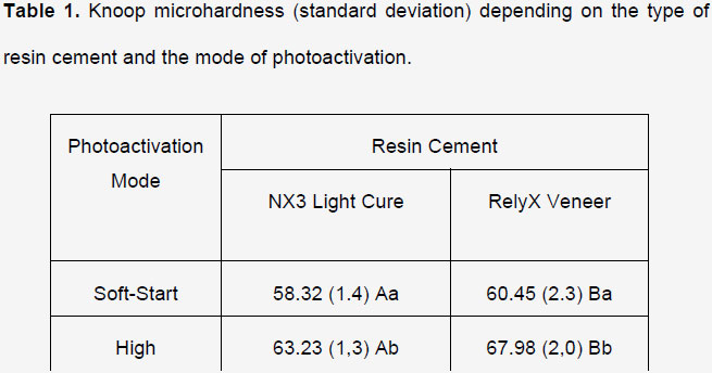
Means followed by distinct letters (horizontal uppercase and vertical lowercase comparing distance within each surface) differ from each other (p≤0.05).
1.4- DISCUSSION
The results of this study showed that, regardless of the mode of photoactivation tested, the RelyX Venner resin cement obtained better performance in relation to the NX3 cement. Therefore, because it is two photopolymerizable resin cements, a possible justification for the superior microhardness of RelyX Veneer cement can be explained by its chemical composition.
When analyzing the composition of the RelyX Veneer cement, this cement is composed of Bis-GMA monomers (bisphenol-A glycidyl methacrylate) and TEGDMA (triethylene glycol dimethacrylate) with zirconia and silica particles18. The presence of TEGDMA reduces cement viscosity significantly increasing the degree of conversion to the polymer. Bis-GMA is capable of promoting strong hydrogen bonding through the hydroxyl group, increasing molecular weight, providing little mobility and high stiffness. There is also a synergistic effect between Bis-GMA and TEGDMA which increases the crosslinking density, that is, crosslinks between linear molecules producing threedimensional polymers with high molecular mass19. In this case, microhardness is an indirect measure of the degree of conversion of monomers into polymers, thus, it can be said that the higher presence of BIS-GMA and TEGDMA provides this cement a higher values of Knoop microhardness, independent of the photoactivation mode. On the other hand, the NX3 Light Cure cement has monomers of uncured methacrylate esters, inert mineral fillers, activators and stabilizers20. The manufacturer does not specify the methacrylate types and does not report the presence of Bis-GMA or TEGDMA, being this a possible hypothesis to justify the lower microhardness values of this cement.
Another factor studied in this study and that deserves to be understood is the difference that occurred for the two resin cements when photoactivated in different ways. The results showed that, regardless of the cement tested, the High Power photoactivation mode showed higher Knoop microhardness values than the Soft-Start mode (gradual increase of light intensity with a reduced contraction voltage and lower temperature). Possibly, this can be explained because in the Soft-Start method the apparatus performs initial cycle of 5 seconds with low irradiance light (average of 601 mW0/cm2) and then reaches a higher irradiance peak (average of 1079 mW/cm2) for 15 seconds. Consequently, when the light was switched on, the apparatus did not reach the maximum intensity of light, the low intensity remaining at a first instant, and only reaching its maximum intensity shortly before the end of the cycle. Thus, the activating effectiveness of photopolymerization occurs only in the repetition of cycles, although the sum of the repeated intensity of the cycles is insufficient compared to the mode of activation of continuous light (High). Therefore, the specimens subjected to the Soft-Start method were subjected to lower light energy rates, causing a lower microhardness14.
1.5- CONCLUSION
From the results found in this study we can conclude that:
Regardless of the photoactivation mode, the RelyX Veneer resin cement presented higher Knoop microhardness when compared to the NX3 Light Cure cement.
High photoactivation mode presented higher Knoop microhardness when compared to Soft-Start mode, independent of resin cement.
1.6- REFERENCES
1. Shetty A.et al. Survival rates of porcelain laminate restoration based on different incisal preparation designs: an analysis. Conserv. J. Dent. 2011; 14:10-5.
2. Strassler HE, Weiner S. Long term clinical evaluation of etched porcelain veneers. J Dent Res. 2001;80:60.
3. Mazaro, J. V. Q. et al. Considerações clínicas para a restauração da região anterior com facetas laminadas. Revista Odontológica de Araçatuba 2009; 30(1): 51-4.
4. El-Badrawy WA, El-Mowafy OM. Chemical versus dual curing of resin inlay cements. J Prosthet Dent.1995; 73:515-24.
5. Pergoraro TA, Silva NR, Carvalho RM.Cements for use in esthetic dentistry. Dent Clin North Am.2007;51(2): 453-71.
6. Arrais CA, Giannini M, Rueggeberg FA. Effect of sodium sulfinate salts on the polymerization characteristics of dual-cured resin cement systems exposed to attenuated light-activation. J Dent. 2009;37(3):219-27.
7. Giorgi MCC, Dias CTS, Paulillo LAMS. Influência das fontes fotoativadorasled e halógena e do tempo de exposição na microdurezaknoop de compósitos odontológicos. Cienc Odontol Bras.2008; 11(4):18-23.
8. Alonso RC, Cunha LG, Correr GM, Cunha Brandt W, Correr-Sobrinho L, Sinhoreti MA. Relationship between bond strength and marginal and internal adaptation of composite restorations photocured by different methods. Acta Odontol Scand.2006;64:306-13.
9. Cunha LG, Alonso RC, Pfeifer CS, Correr-Sobrinho L, Ferracane JL, Sinhoreti MA. Modulated photoactivation methods: Influence on contraction stress, degree of conversion and push-out bond strength of composite restoratives. J Dent. 2007; 35(4):318-24.
10. Baratieri LN, Monteiro SJ. Odontologia Restauradora: Fundamentos & Técnicas. São Paulo: Santos; 2012. Volume 1.
11. Santos MJMC, Silva e Souza JR MH, Mondelli RFL. Novos conceitos relacionados à fotopolimerização das resinas compostas. JBD.2002;1(1):14-21.
12. Cunha LG, Alonso RC, Correr GM, Brandt WC, Correr-Sobrinho L, Sinhoreti MA. Effect of different photoactivation methods on the bond strength of composite resin restorations by push-out test. Quintessence Int. 2008 Mar;39(3):243-9.
13. Alonso RC, Cunha LG, Correr GM, Cunha Brandt W, Correr-Sobrinho L, Sinhoreti MA. Relationship between bond strength and marginal and internal adaptation of composite restorations photocured by different methods. Acta Odontol Scand 2006;64:306-13.
14. Consani S, Pereira SB, Sonhoreti MA, Correr Sobrinho L.Effect of the methods of photoactivation and insertion on the hardness of composite resins. Pesqui Odontol Bras.2002; 16(4): 355-60.
15. Nomoto R, Uchida K, Hirasawa T. Effect of light intensity on polymerization of light-cured composite resins. Dent Mater J 1994;13:198-205.
16. Price RB, Felix CA, Andreou P. Knoop hardness of ten resin composites irradiated with high-power LED and quartz-tungsten-halogen lights. Biomaterials.2005;26(15):2631 -41.
17. Borges AB, Yui KCK, D'avila TC, Takahashi CL, Torres CRG, Borges LS. Influence of Remineralizing Gels on Bleached Enamel Microhardness In Different Time Intervals. Operative Dentistry.2010;35(2):180-6.
18. Leal CL, Queiroz APV, Foxton RM, Argolo S, Mathias P, Cavalcanti AN.Water Sorption and Solubility of Luting Agents Used Under Ceramic Laminates With Different Degrees of Translucency. Oper Dent.2016; 41(5):141-8.
19. Gonçalves F, Kawano Y, Pfeifer C, Stansbury JW, Braga RR. Influence of BisGMA, TEGDMA, and BisEMA contents on viscosity, conversion, and flexural strength of experimental resins and composites. Eur J Oral Sci. 2009; 117(4):442-6.
20. Ishikiriama SK, Ordoñéz-Aguilera JF, Maenosono RM, Volú FL, Mondelli RF.Surface roughness and wear of resin cements after toothbrush abrasion. Braz Oral Res. 2015; 29:1-5.
1. Student of the Dentistry Course at Faculty of Medical Sciences and Health of Juiz de Fora - Suprema. - Student 2. Student of the Dentistry Course - Student 3. PhD in Dental Clinics - Restorative Dentistry at FOP/UNICAMP - Adjunct Professor of the Dentistry Course at FCMS/Suprema