
INTRODUCTION
Particulate matter (PM) emitted by diesel exhaust (DEP) comprise a mixture of gases, vapors, organic compounds and fine particles released from vehicle engineering. Considered one of the main villains in triggering inflammatory and allergic reactions, since their fine particles (PM <2.5 µm penetrate more easily through the airways, possibly causing pulmonary complications (1). Biochemical evidence demonstrate that toxic chemical molecules such as quinones (1,2-NQ) are incorporated into the surface of the PED, making them more toxic (2). Considered a physical barrier and metabolically active, the lung epithelium contributes to the maintenance of homeostasis in the airways. According to Davies et al.,(3) inflammation and airway remodeling appear to relate to the susceptibility of the bronchial epithelium of patients to inhaled environmental agents. Airways contain abundant innervation of sensory afferent fibers(4); stimulation of these fibers by chemical mediators is modulated by specific receptors, among them the vanilloid receptor-1 (TRPV1), known to be activated by capsaicin, a pungent ingredient extracted from red peppers of the genus Capsicum(5). Administration of capsaicin to newborn rats (2-10 days old) at a dose of 50 mg/kg, promotes maximum degeneration of unmyelinated sensory afferent fibers with consequent neurochemical, histochemical and functional impairment (6). Connections exist between pollutants and vascular disorders (e.g. inflammation) via mechanisms modulated by oxidative stress and NO (1), cytokines such as TNF-α which has biological effects mediated by interaction with its cell surface receptors: TNFR-I and TNFR-II (7); activation of transcription factor NF- κ B in epithelial cells of the respiratory tract (8), but little is known about the association between environmental pollutants and the involvement of neuro-inflammatory components (e.g. neuropeptides).
2. OBJECTIVE
To study the relationship of inflammation promoted by environmental pollutants: PED and 1,2-NQ with neurogenic mechanisms in the main bronchus of the rat.
2 METHODS 2.1 Animals
The project was developed according to the rules governed by the Animal Ethics Committee of the Institute of Biomedical Sciences, Universidade de São Paulo. Wistar rats of both sexes (250 - 350g) and, in certain protocols, newborn Wistar rats (2-10 days old) treated with capsaicin at 50 mg/kg, were used. The animals received intraperitoneal (ip) anesthesia with a mixture of ketamine (80 mg/kg) and xylazine (16 mg/kg). After induction of anesthesia, they received intratracheal (i.tr.) injection of test agents and were sacrificed after 3 hours.
2.2 Quantification of gene expression (TNF- α, TNFR1, TNFR2, NF- κ B, NOSi) by real time
The animals were treated with pollutants and three hours later the main bronchi were analyzed for gene expression of TNF-α and its receptors: R1 and R2, as well as NF-α B and iNOS by PCR assay. Total RNA extraction was performed by the Trizol method. Real-time PCR is based on the presence of fluorophore, present in the PCR buffer, which releases fluorescence proportional to the number of DNA copies.
2.3 Statistical analysis
They were subjected to one-way analysis of variance (ANOVA) followed by modified Bonferroni's test. Differences between means were evaluated with the help of a statistical program (Prism 4.0 software). P values <0.05 were considered significant.
3. RESULTS 3.1 Quantification of gene expression of TNF-α , TNFR1, TNFR2, NF- κ B and iNOS by real time PCR in the main bronchus of rats exposed to pollutants
The gene expression of cytokine TNF- α and its receptors TNFR1 and TNFR2 was increased in bronchus from rats treated with 1,2-NQ alone compared with its vehicle (Figure 1A-C). In the bronchus of rats treated with DEP, expression of the TNFR1 receptor, but not TNF- α and the TNFR2 receptor, was affected (P <0.05) when compared with animals that received vehicle (Figure 1A-C). In the group of animals that received the mixture of pollutants (DEP + 1,2-NQ), a significant increase was observed only in the expression of the TNFR1 receptor (P <0.001) compared with the vehicle group.
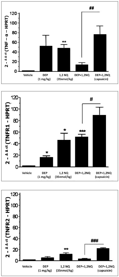 Figure 1 A, B, C. Gene expression analysis of TNF- α(panel 1A) and its receptors TNFR1 (panel 1B), TNFR2 (panel 1B), TNFR2 (panel 1C) in the main bronchus of rats. Data are expressed as mean ± SEM for n = 3-5 animals. * P <0.05, ** P <0.01 and *** P <0.001 versus vehicle . # P <0.05, # # P <0.01 and # # # P <0.001 versus the group treated with the mixture of pollutants that received capsaicin when neonates.
In the group of animals pretreated with capsaicin that received the mixture of pollutants a significant increase in gene expressions of TNF-α (P <0.01) and its receptors TNFR1 (P <0.05) and TNFR2 (P < 0.001) was observed when compared with the group of animals that received only the mixture of pollutants (Figure 1A-C).
No significant changes were observed in gene expression of NF-kB in the different groups (Figure 2).
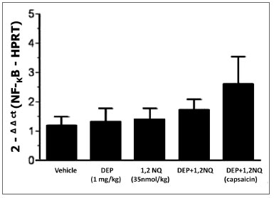 Figure 2. Gene expression analysis of transcription factor NF-kB in the main bronchus of rats. Data are expressed as mean ± SEM for n = 3-5 animals.
The mixture of pollutants caused no significant change in the expression of iNOS, except with PED alone (P <0.05) compared to the vehicle group. However, in animals treated with capsaicin, the administration of the pollutant mixture caused a marked increase in the expression of iNOS compared to animals exposed to the same pollutants which had not been treated with capsaicin (P <0.01, Figure 3).
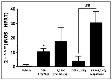 Figure 3. Gene expression analysis of iNOS in the main bronchus of rats treated i.tr. with pollutants and their vehicles (after 3 h). Data are expressed as mean ± SEM for n = 3-5 animals. * P <0.05 versus vehicle. # # P <0.01 versus the group treated with the mixture of pollutants that received capsaicin when neonates.
4. DISCUSSION
Considered one of the major components of the PM environment in urban areas, DEP have been implicated in triggering inflammatory diseases of the airways (9). In addition, reactive chemical compounds such as quinones and their reduction products (e.g. hydroquinone, semiquinones), found in nature or in PM suspended in air (2), are of particular toxicological interest, because they produce, via complex mechanisms, a variety of harmful effects on the health of the population. Among these effects are: the ability to generate oxygen-reactive species, promoting covalent bonds with tissue macromolecules (10) that lead to cytotoxicity and cell death (2) and also interfere with enzymatic reactions, causing immunotoxicity and carcinogenesis (11).
The inflammation process of the airways is associated with increased production of proinflammatory cytokines by epithelial cells (12), macrophages (13), mast cells (14) and eosinophils (15). Cytokines also have an important role in cell activation, proliferation, cell differentiation, adhesion, migration and synthesis of acute phase proteins. In addition, clinical and experimental studies suggest cytokines are a target in chronic obstructive pulmonary diseases (16). Among cytokines involved in inflammatory reactions of the airways, TNF- α has a leading role, because in addition to recruiting leukocytes into the airways, it increases the adhesion of neutrophils and monocytes to the cellular matrix, stimulates fibroblast proliferation and production of other inflammatory cytokines (17).
It is interesting to note that the proinflammatory cytokine TNF- α has stimulant action on nuclear factors, including factor NF-kB (18). Despite high levels of this cytokine in bronchial homogenates of rats exposed to pollutants, real-time PCR analysis did not detect significant increase in the concentration of the protein NF-kB in the bronchi of the animals, indicating that at the time point investigated, the concentration of TNF-α formed did not interfere in the generation of factor NF-kB.
Besides cytokines, the involvement of reactive species (NO) in the inflammatory response has been amply demonstrated in the literature. This product was also found in atmospheric pollution and cigarette smoke (19). In general, the cytotoxicity of NO is related to the molecule's action with other compounds released during inflammation, and this study's PCR assay showed significant increase in gene expression of iNOS in the main bronchus of rats treated i.tr. with DEP and with the mixture of pollutants, pre-treated with capsaicin but not in animals treated with 1,2-NQ or a mixture thereof, which did not receive capsaicin.
Airways are densely innervated by sensory nerve fibers (20) which, by appropriate stimuli, both endogenous (e.g. chemical mediators of inflammation) or exogenous (e.g. capsaicin, microorganisms), culminate in the release of neuropeptides, capable of initiate responses/defense mechanisms such as cough, local vasodilation, bronchoconstriction and mucus secretion. These can be classically defined as neurogenic inflammation. The inflammatory results obtained in rats treated with capsaicin (neuropeptide-depleted) were potentiated when compared with healthy animals. Although intriguing, this finding corroborates other literature data, which suggest that the degeneration of sensory fibers (type C), by capsaicin, exacerbated the inflammatory response in several experimental models.
Among the studies, Medeiros et al.,(21) showed that neonatal treatment of rats with capsaicin increased neutrophilia evoked by OVA in bronchoalveolar lavage and pleural cavity. This same treatment promoted hyperreactivity in rat airways(22). In the animals, treatment with capsaicin contributed to a greater increase in bronchial reactivity in response to cigarette smoke (23).
The pooled data obtained in this study together with those described in the literature, indicate that the neuropeptides biosynthesis impairment, especially tachykinins, is a very relevant aspect for the positive regulation of neutropoiesis. It is likely that the increased inflammatory response induced by DEP and 1,2-NQ in the bronchi of animals treated with capsaicin is partly due to the increase in pro-inflammatory cytokines genesis. The significant increase in gene expressions of TNF- α and its receptors (TNFR1 and TNFR2) in reaction to the pollutants in the bronchus of rats pretreated with capsaicin compared to healthy animals reinforces this hypothesis.
Additionally, two other hypotheses can be discussed to better explain the increased influx of neutrophils in reaction to pollutants in neuropeptides-depleted animals: the first, based on evidences by Franco et al., (24) which suggest that the permanent loss of C fibers and the consequent suppression of neuropeptides production by treating neonate rats with capsaicin leads to increased expression/regulation of mast cell lung residents. Thus, we can postulate that if mast cells are present in greater quantities in the lung parenchyma of rats treated with capsaicin, that the rats, when stimulated by the mixture of pollutants, will result in increased cytokine production and consequently in greater leukocytes influx, therefore the local neutrophil influx induced by pollutants in the bronchi of neuropeptides-depleted rats occurred as a result of increased gene regulation of some cytokines by mast cells.
In conclusion, the results presented in this study allow us to suggest that the increase in the production and/or release of endogenous inflammatory mediators such as cytokines, greatly modulates the inflammatory response induced by environmental pollutants DEP and 1,2-NQ. Their autopharmaclogical process is intensified in animals deprived of sensory nerve fibers. This is due partly to the fact that in these animals, the absence of neuropeptides (possibly tachykinins) resulted in increased macrophage functional activity, coupled with the production of proinflammatory cytokines by these cells and possibly by mast cell lung residents. These evidences contribute to a growing argument that the loss of tachykinergic afferent signals leads to worsening of the inflammation induced by irritants, including the pollutants DEO and 1,2-NQ.
5. ACKNOWLEDGEMENTS
Pollutants were kindly provided by Prof. Dr. Yoshito Kumagai (University of Tsukuba, Japan) and the Wistar rats treated with capsaicin by Prof. Dr. Edson Antunes (Universidade de Campinas). We also thank the technical support of Mrs. Maria Alice Barreto.
6. REFERENCES
1. Donaldson K. The biological effects of coarse and fine particulate matter. Occup Environ Med 2003 May;60(5):313-4.
2. Cho AK, Schimitz DA, You Y, Rodriquez CE, Distefano E, Kumagai Y, et al. Detemination of four quinones in Diesel exaust particles, SRM1649A and athmosferic 2.5. Aerosol Sci Technol 2004;38:1 -14.
3. Davies DE, Holgate ST. Asthma: the importance of epithelial mesenchymal communication in pathogenesis. Inflammation and the airway epithelium in asthma. Int J Biochem Cell Biol 2002 Dec;34(12):1520-6.
4. Joos GF, Germonpre PR, Pauwels RA. Role of tachykinins in asthma. Allergy 2000 Apr;55(4):321-37.
5. Caterina MJ, Schumacher MA, Tominaga M, Rosen TA, Levine JD, Julius D. The capsaicin receptor: a heat-activated ion channel in the pain pathway. Nature 1997 Oct 23;389(6653):816-24.
6. Jancso N, Jancso-Gabor A, Szolcsanyi J. Direct evidence for neurogenic inflammation and its prevention by denervation and by pretreatment with capsaicin. Br J Pharmacol Chemother 1967 Sep;31(1):138-51.
7. Wallach D, Varfolomeev EE, Malinin NL, Goltsev YV, Kovalenko AV, Boldin MP. Tumor necrosis factor receptor and Fas signaling mechanisms. Annu Rev Immunol 1999;17:331-67.
8. Dagher Z, Garcon G, Billet S, Verdin A, Ledoux F, Courcot D, et al. Role of nuclear factor-kappa B activation in the adverse effects induced by air pollution particulate matter (PM2.5) in human epithelial lung cells (L132) in culture. J Appl Toxicol 2007 May-Jun;27(3):284-90.
9. Nemmar A, Al-Maskari S, Ali BH, Al-Amri IS. Cardiovascular and lung inflammatory effects induced by systemically administered diesel exhaust particles in rats. Am J Physiol Lung Cell Mol Physiol 2007 Mar;292(3):L664-70.
10. Cadenas E, Hochstein P, Ernster L. Pro- and antioxidant functions of quinones and quinone reductases in mammalian cells. Adv Enzymol Relat Areas Mol Biol 1992;65:97-146.
11. Monks TJ, Jones DC. The metabolism and toxicity of quinones, quinonimines, quinone methides, and quinone-thioethers. Curr Drug Metab 2002 Aug;3(4):425-38.
12. Ackerman V, Marini M, Vittori E, Bellini A, Vassali G, Mattoli S. Detection of cytokines and their cell sources in bronchial biopsy specimens from asthmatic patients. Relationship to atopic status, symptoms, and level of airway hyperresponsiveness. Chest 1994 Mar;105(3):687-96.
13. Gosset P, Tsicopoulos A, Wallaert B, Joseph M, Capron A, Tonnel AB. Tumor necrosis factor alpha and interleukin-6 production by human mononuclear phagocytes from allergic asthmatics after IgE-dependent stimulation. Am Rev Respir Dis 1992 Sep;146(3):768-74.
14. Gordon JR, Burd PR, Galli SJ. Mast cells as a source of multifunctional cytokines. Immunol Today 1990 Dec;11(12):458-64.
15. Shah A, Church MK, Holgate ST. Tumour necrosis factor alpha: a potential mediator of asthma. Clin Exp Allergy 1995 Nov;25(11):1038-44.
16. Chung KF. Cytokines as targets in chronic obstructive pulmonary disease. Curr Drug Targets 2006 Jun;7(6):675-81.
17. Thommesen L, Sjursen W, Gasvik K, Hanssen W, Brekke OL, Skattebol L, et al. Selective inhibitors of cytosolic or secretory phospholipase A2 block TNF-induced activation of transcription factor nuclear factor-kappa B and expression of ICAM-1. J Immunol 1998 Oct 1;161(7):3421-30.
18. Solt LA, Madge LA, Orange JS, May MJ. Interleukin-1-induced NF-kappaB activation is NEMO-dependent but does not require IKKbeta. J Biol Chem 2007 Mar 23;282(12):8724-33.
19. Borland C, Higenbottam T. Nitric oxide yields of contemporary UK, US and French cigarettes. Int J Epidemiol 1987 Mar;16(1):31-4.
20. Spina D, Page CP. Pharmacology of airway irritability. Curr Opin Pharmacol 2002 Jun;2(3):264-72.
21. Medeiros MV, Macedo-Soares MF, De Luca IM, Hyslop S, De Nucci G, Antunes E. Contribution of C-fibers to leucocyte recruitment in bronchoalveolar lavage fluid and pleural cavity in the rat. Eur J Pharmacol 2001 Jun 8;421(2):133-40.
22. Medeiros MV, De Luca IM, Bricola AA, Zanesco A, De Nucci G, Antunes E. Enhanced airways responsiveness in rats depleted of sensory neuropeptides by neonatal capsaicin treatment. Neurosci Lett 2003 May 1;341(2):103-6.
23. Mutoh T, Joad JP, Bonham AC. Chronic passive cigarette smoke exposure augments bronchopulmonary C-fibre inputs to nucleus tractus solitarii neurones and reflex output in young guinea-pigs. J Physiol 2000 Feb 15;523 Pt 1:223-33.
24. Franco-Penteado CF, De Souza IA, Camargo EA, Teixeira SA, Muscara MN, De Nucci G, et al. Mechanisms involved in the enhancement of allergic airways neutrophil influx by permanent C-fiber degeneration in rats. J Pharmacol Exp Ther 2005 Apr;313(1):440-8.
1. Ph.D. (Researcher). 2. Ph.D (Professor at the School of Arts, Sciences and Humanities (EACH) at Universidade de São Paulo). 3. Tenured professor (Associate Professor, Department of Pharmacology, Institute of Biomedical Sciences, Universidade de São Paulo).
Correspondence: Teles AM Rua Dr. Pena Forte Mendes, 86 Apart. 13. Bela Vista São Paulo-SP Cep. 01308-010 Phone: (11) 3214-0597 E-mail address: tmausp@yahoo.com
This article was submitted to SGP (Publication Management System) of Rev Bras Cien Med Health on March 1, 2009. Cod. 11. Accepted on October 2, 2009. CNPq; FAPESP