
INTRODUCTION
Currently, physical therapy has been on the spotlight due to the benefits it produces in a wide variety of musculoskeletal dysfunctions, among them craniomandibular disorders (CMD). Its prevalence has considerably increased, with its signs and symptoms affecting 86% of Western populations.(1) Although it can occur at any age, is more common among individuals of 13-35 years of age. This disorder is four times more prevalent in women than in men.(2)
CMD is defined as a pathological condition affecting the temporomandibular joint (TMJ), muscles of mastication and adjacent components.(3) The greatest cause of pain in CMD is musculoskeletal, and involves the neck and the muscles of mastication. It has a multifactor etiology, and many studies seek to define its predisposing, causal and perpetuating mechanisms.(3,4,5,6,7) Classic symptoms of CMD are noises in the mandible, limitation of movements and/or deviations of jaw movements, pain in the temporomandibular joint and/or mastication muscles and cervical area.(4,6,8,9,10, 11, 12)
Physical therapy treatment aims to relieve osteomuscle pain, reduce inflammation and restore oral motor function. The most recommended physical therapies to control CMD are therapeutic exercise and manual therapy techniques.(7)
Among the various manual therapy techniques, Mulligan's stands out. This technique was developed by physical therapist Brian Mulligan in 1954, in New Zealand. Mulligan had the cooperation of the leading international exponents of manual therapy, such as Maitland, Cyriax, Elvey, and Mackenzie.(4)
Since 1972, Brian Mulligan and his team have diffused his technique throughout the world. It is based on the theory of positional fault, that says that when there is an injury, the joint can take a slightly unusual position, often invisible in examinations such as X-rays, and that these small faults cause restrictions of movement with or without pain.(4)
When accessory movements are associated with active free movements they are called SNAGS (sustained natural apophyseal glides). When only accessory movements are used, they are called NAGS (natural apophyseal glides). It is expected that immediately after the applying the technique the function is increased and the pain is decreased.(13)
Both the SNAGS and NAGS are only applied to the spine. Movements used in the extremities are called MWM (mobilizations with movements).(4). In this study we only used the first two techniques.
METHODS
Type of study: original study
Study site: Hospital e Maternidade Theresinha de Jesus at the Physical Therapy Clinic School.
Eligibility criteria: patients and intern student at the Physical Therapy Clinic School.
Inclusion criteria: patients with craniomandibular dysfunction.
Exclusion criteria: Patients who made use of analgesics and/or muscle relaxant up to 8 hours before the physical therapy procedure and that showed hypermobility in the cervical region or mouth opening.
Sample and procedures: fifteen subjects with craniomandibular dysfunction were evaluated in the study, one female patient was excluded for presenting hypermobility of mouth opening. Fourteen individuals participated in the study, three men and eleven women. First, the patients underwent a standard assessment, which consisted of a history to record general patient data, a osteokinematic mobility test of the cervical spine, cervical spine and TMJ pain assessment by visual analog scale of pain. Initially, mouth opening was evaluated with a ruler placed between the upper and the lower central incisors, the reference value was 3.5 to 5.0 cm.(14) If the patient presented hypermobility of mouth opening or cervical spine he/she was excluded from the study. The four movements of the cervical spine were evaluated. To evaluate flexion, the patient was requested to perform the movement and then the distance between the bone prominence of the chin to the sternal notch was measured with a tape. To evaluate extension, first, the patient was requested to perform the movement and then the distance between the bone prominence of the chin to sternal notch was measured with a tape. To assess rotation, the patient was requested to perform the movement and then the distance between the bony prominence of the chin and the distal bone prominence of the acromion was measured with a tape. To evaluate lateral inclination the distance between the proximal bone prominence mastoid process of temporal bone and the distal bone prominence of the acromion is measured. To evaluate cervical spine movements, the patient will remain seated with his/her mouth closed.(15. The following parameters were considered for normal ROM: flexion 2.5 cm, extension 17.5 to 25 cm, 12.5 cm, rotation and lateral inclination 12.5 cm.(14) Cervical pain and TMJ pain was assessed by visual analog scale of pain. This scale consists of a 10-centimeters long line, drawn on a paper, in which the patient indicates with a mark with a pen, the point where the pain is located at that moment.(16) The therapist first explains to the patient that the start of the scale is equivalent to no pain and the end equals the unbearable pain. Without being noticed by the patient, the therapist assigns a value between zero (0) and ten (10) to the mark, where 0 = no pain, 10 = unbearable pain. After initial evaluation, patients were submitted to the SNAGS and NAGS techniques C3 to C5. The first, SNAGS technique, consists of a sliding device applied to a vertebral segment with an associated physiological active movement, which is sustained at the beginning and end of the motion. The second technique, NAGS, consists of passive accessory movements applied in the cervical and upper thoracic spine (13). Then, a second evaluation, identical to the initial evaluation, is performed to determine whether there was change in pain and range of motion of the cervical spine and temporomandibular joint. The study was approved by the Ethics Committee of the Medical Sciences and Health School of Juiz de Fora, and all patients signed an informed consent.
RESULTS
Initially, testing was performed to investigate whether the variables showed normal distribution. A t test was performed for variables with normal distribution and a Mann Whitney test was used for outliers.
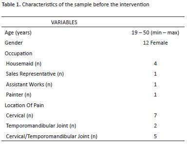
In table 1, there are sample characteristics and location of pain before intervention.
The only variable that did not achieve statistical significance was cervical flexion ROM, p = 1.000 (Figure 1).
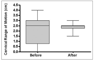 Figure 1. Cervical flexion RM p = 1.000
Cervical mobility showed a significant improvement in relation to cervical extension movements p = 0.000 (Figure 2), right lateral inclination p = 0.001 (Figure 3), left lateral inclination p = 0.005 (Figure 4), right rotation p = 0.002 (Figure 5), left rotation p = 0.003 (Figure 6), neck pain p = 0.004 (Figure 7), TMJ pain p = 0.041 (Figure 8), mouth opening p = 0.001 (Figure 9).
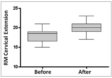 Figure 2. Cervical extension RM p = 0.000
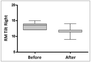 Figure 3. Tilt Right RM p=0.001
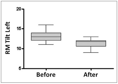 Figure 4. Tilt Left RM p = 0.005
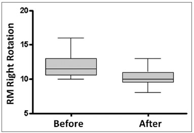 Figure 5. Right Rotation RM p=0,002
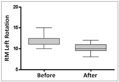 Figure 6. Left Rotation RM p=0,003
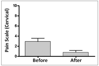 Figure 7. Cervical Pain Scale p=0,004
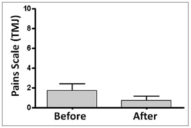 Figure 8. Pain Scale TJM p=0,041
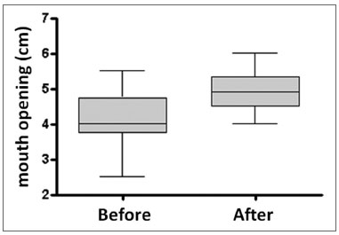 Figure 9. Mouth opening p=0,001
DISCUSSION
The study results were statistically significant for all variables, except for cervical flexion. This can be explained by the fact that the average values of flexion before (2.0 ± 1.3 cm) and after ( 2.3 ± 0.4 cm) were close to normal values, 2.5 cm.
Regarding the variable TMJ pain, the p value was near 0.05. This may have occurred because the therapeutic technique was applied in the cervical region and not directly at the TMJ, as the study's objective was to assess to which extent improvement in neck pain influences TMJ pain.
The posture of each individual is determined by muscular chains, fascia, ligaments and bone structures, which are interlinked and cover the whole body.(18) Every time a muscle shortens, its extremities get nearer each other and there is a shift on the bones on which it is inserted, so the joints are blocked and the body is deformed. Therefore, all other muscles that insert on the bone will be affected by the displacement, which will propagate to other bones and muscles, and so on. Several studies have shown that patients with CMD present changes in the position of the head and shoulders, as well as increased or rectification of cervical lordosis.(18,19)
Some studies suggest that when anteriorization of the head occurs, the gaze is lowered and in an attempt to level the gaze there is an increase in cervical lordosis, reflecting on the muscles of mastication.(19) Other authors explain that because mastication muscles are synergistic to cervical muscles, any imbalance between them causes retrusive forces in the jaw, altering its rest position and leading to muscle hyperactivity.(20)
According Amantéa et al.,(1) the balance of the body and head movements are generated by the positioning of the skull over the cervical area, thereby determining the individual's posture. The temporomandibular joint (TMJ) is directly related to cervical and scapular areas through a common neuromuscular system, so postural changes in the cervical spine can result in CMD and vice versa.
We observed that half of the participants in the study were students, and this may have influenced results, as student posture disfavors normal biomechanics of the TMJ. Individuals who work as painters or builders also have a significant disadvantage in cervical spine and TMJ biomechanics. Usually individuals who work in these professions are unaware of the proper biomechanics for conducting their work activities.
The real cause of TMJ dysfunction in the sales promoter is not known. It may occur due to a dental malocclusion due to genetic causes. The muscles must work in a coordinated manner and for this the occlusion must be balanced.(21) Malocclusion may lead to muscle spasms and a temporomandibular disorder, or the tension in the neck caused by the profession due to targets to be achieved and the fact that this profession requires a lot of verbal communication, overloading the TMJ.(22)
Domestic work differs from other professions mentioned above because the movements are varied, highlighting the use of muscles in the upper limbs, often asymmetrically. Domestic activities involve movements while sitting or standing, and require pushing, pulling and lifting weights.(23) According to Dulweerdmeester (1998), environmental factors such as noise, vibration, lighting, climate, chemical agents, information captured by vision, hearing and other senses also influence the ergonomics of housework.
According to Stenks et al. (6) TMJ dysfunctions involve cranio-cervical-mandibular skeletal components and its relationship with teeth and the neuromuscular system. The lack of harmony of the teeth, TMJ and neuromuscular system favors the emergence of myofascial dysfunction. Muscle dysfunction is the most common clinical diagnosis, however, its symptoms are complex, with variations directly related to musculoskeletal and behavioral factors.(4)
Physical therapy in TMD aims to relieve osteomuscle pain, reduce inflammation, restore oral motor function and cervical mobility and decrease pain.(4)
This study agrees with Marenga S et al. (11), who in their systematic review of physical therapy in TMD, concluded that manual mobilizations may be effective in the short-term to increase mouth opening in TMD patients. This author suggests that laser therapy may decrease pain and improve mouth opening and may be more effective than other electrotherapeutic resources in the short term, but could not say whether lasertherapy is more effective compared to manual therapy to improve the TMJ function.(24,25)
The various forms of physical therapy interventions have demonstrated good results in the treatment of TMD. Nikolis et. al. studied the effect of active and passive movement exercises in the TMJ and global posture correction in TMD and, at the end of the study, found that interventions reduced pain, concluding that therapy through exercises may be useful in the treatment of TMJ dysfunction.(26) Wright et al. Noted that posture training along with instructions to move the TMJ are effective for individuals with TMD and that posture training has a positive impact on reducing TMD symptoms.(27)
The study by Nicolakis et al. used exercise and manual therapy for TMD patients and achieved a success rate of 90% reduction in TMJ pain and increase in mouth opening in short-term treatment. The study concluded, based on these findings, that exercise therapy is an effective tool for treating TMD.(28) These results corroborate ours in regard to mouth opening and pain reduction, but our results were only related to manual therapy.
Taylor M et al. observed that TMD is characterized by reduced mandibular movement and increased activity of the masseter muscle and showed that individuals with significant decrease in masseter activity also showed increased mobility of the TMJ. Their results suggested that mobilization of the TMJ is an effective way to reduce muscle tension, and in the short run, increase the range of mandibular movement.(29) Our study agrees with this as regards the reduction of movement of mouth opening in TMD patients. We also observed that the tested individuals had pain and reduced movement of the neck. Our results demonstrate that manual therapy is an effective treatment in the short term, to increase the mobility of the jaw, but our intervention was in the cervical area, while the quoted study acted on the TMJ.
Despite the positive results found in this study, in which we evaluated manual techniques in the short term, we see the need for more studies evaluating the same techniques in the long-term and involving a control group, randomization of the sample and blinding of the assessor and the patient.
CONCLUSION
Mulligan's technique for cervical mobilization can be effective in the short term to increase cervical and mouth opening range of motion, and can reduce pain in the TMJ and cervical spine.
REFERENCES
1. Amantea DV, Novaes AP, Campolongo GD, et al. A importância da avaliação postural no paciente com disfunção da articulação temporomandibular. Acta ortop Brás 2004;12(3):155-159.
2. Bove SRK, Guimarães AS, Smith RL. Caracterização dos pacientes de um ambulatório de disfunção temporomandibular e dor orofacial. Rev Latino Am Enfermagem 2005;13(5):686-691.
3. Moana Filho EJ. Levantamento das atitudes e crenças dos ortodontistas com relação à disfunção têmporo-mandibular e dor orofacial. Rev Dent Press Ortodon Ortop Facial 2005;10(4):60-75.
4. Oliveira AS de, Bermudez CC, Souza RA, et al. Impacto da dor na vida de portadores de disfunções temporomandibular. J Appl Oral Sci 2003;11(2):138-43.
5. Pedroni CR, Oliveira AS, Bérzen F. Paint characterísticas of temporomandibular disorder: A pilot study in patients with cervical spine dysfunction. J Appl Oral Sci 2006;14(5):388-92.
6. Stenks DH, Wijer A. Disfunções da articulação temporomandibular do ponto de vista da fisioterapia e da odontologia. São Paulo: Ed. Santos;1996.
7. Santos ACE, Bertoz AF, Pignatta BML, et al. Avaliação Clínica de sinais e sintomas da disfunção temporomandibular em crianças. R Dental Press Ortodon Ortop Facial 2006;11(2):29-34.
8. Pereira FNK, Andrade LLS de, Costa MLG. Sinais e sintomas de pacientes com disfunção temporomandibular. Rev Cefac 2005;7(2):221-8.
9. Neely MLC, Olivo SA, Magge DJ. A Systematic Review of the effectiveness of physical therapy interventions for temporomandibular disordens. Physical Therapy 2006;86(5):710-725.
10. Ilha VCA, Et al. Estimativa da excursão condilar em pacientes com disfunção craniomandibular:um enfoque multidisciplinar. Rev Press Ortodon Ortop Facial 2006;11(3):63-70.
11. Rodrigues CPC, Silva MJE, Flávia O. Ruídos articulares e sinais de disfunção temporomandibular: um estudo comparativo por meio de palpação manual e vibratografia computadorizada da ATM. Pesq Odont Brás 2000;14(4): 367-371.
12. Bruno, MAD. Disfunção têmporo-mandibular: aspectos clínicos de interesse do cefaliatra. Migrâneas cefaléias. Rev Press Ortodon Ortop Facial 2004;7(1):14-18.
13. Tidswell, Marian. Ortopedia para fisioterapeutas. São Paulo: Editorial Premier; 2001.
14. Magee, DJ. Avaliação Musculoesquelética. São Paulo: Ed. Manole; 2005.
15. M. Linn Palmer, Marcia E.Epler. Fundamentos das técnicas de avaliação musculoesquelética. São Paulo: Ed.Guanabara koogan; 2000.
16. Viel E. O Diagnóstico Cinesioterapêutico. São Paulo: Ed.Manole; 2001.
17. Pereira Junior FB, Tunes RS, Silva ALR. Anquilose da articulação temporo-mandibular causada por agressão por arma de fogo: Relato de Caso. Rer Cir Traumatol Buco-maxilo-fac 2005:5(4):37-42.
18. Wright EF, Domenech M, Fischer JR. Utilidade do treinamento da postura para pacientes com desordens temporomandibular. Dente Assoc de J Am 2000;131:202-210.
19. Burgess JA, Sommers EE, Truelove EL, Dworkin SF. Efeitos a curto prazo de dois métodos terapêuticos na dor miofascial e deficiência orgânica do sistema mastigatório. Dente de J Prosthet 1998;60:606-610.
20. Yi LC, Guedes ZCF, Vieira MM. Relação da postura corporal com a disfunção da articulação temporomandibular: Hiperatividade dos músculos da mastigação. Fisioterapia Brasil 2003;4(5):341-347.
21. Darling DW, Krauss S, Clasheen-Wray MB. Relationship of head posture and the rest position of the mandible. J Prost Dent 1994;52(1):111-15.
22. Sousa LE, et. Al. Relação entre o estresse e as disfunções da ATM nos alunos e professores da Universidade Católica de Petrópolis. Fisioterapia Brasil 2004;5(5):363-368.
23. Dul J,Weerdmeester B. Ergonomia Prática. São Paulo: Ed. Edgard Bluncher; 1998.
24. Marenga S Medlicott, SUSAN R Harris. A Systematic Review of the effectiveness of exercise, manual therapy, electrotherapy, relaxation training, and biofeedback in the management of temporomandibular disorder. Physical Therapy 2006;86(7):955-973.
25. Bjordal JM, Couppe C, Comida RT. Uma revisão sistemática da terapia de baixo nível do laser com doses posição-específicas para a dor das desordens crônicas. Aust J Physiother 2003;49:107-116.
26. Nikolis, P. et al. Effectiveness of Exercise Therapy in Patients with Myofascial Pain Dysfunction Syndrome. Journal of Oral Rehabilitation 2002;29:362-368.
27. Wright EF, Domenech M, Fischer JR. Utilidade do treinamento da postura para pacientes com desordens temporomandibular. Dente Assoc de J Am 2000;131:202-210.
28. Nicolakis P, Erdogmus CB, Kollmitzer J, et al. Long-term outcome after treatment of temporomandibular joint osteoarthritis with exercise and manual therapy. Cranio 2002;20(1):23-27.
29. Taylor M, Suvinen T, Reade P. The effect of Grade IV distraction mobilization on patients with temporomandibular pain-dysfunction disorder. Physiotherapy Theory and Practice 1994;10:129-136.
1 Graduation (Physical therapist). 2 Graduate (Physical Therapist). 3 Graduation (Physical Therapist). 4 MSc (Professor). 5 MSc (Professor).
Faculdade de Ciências Médicas e da Saúde de Juiz de Fora (Medical Sciences and Health School of Juiz de Fora)
Taís Resende Di Mambro taisrdm@gmail.com
This article was submitted to SGP (Publication Management System) of Rev Bras Cien Med Health on March 26, 2009. Cod. 15. Accepted on October 2, 2009.