
INTRODUCTION
Auriculotherapy is a technique that has been used for thousands of years; it is a system to diagnose and treat physical and psychosomatic dysfunctions, based on the normalization of the organism (1-4). It is also used as an alternative treatment, where a specific point is stimulated in the auricular pavilion represented by the external ear that connects with the whole human body (4-8) and involves reflex neurological pathways, neurotransmitters, cytokines, immune system, among others. This technique, which is part of Traditional Chinese Medicine (TCM), is based on the principle that there is a harmonization of the obstructed meridian and that the ear is directly or indirectly connected with twelve of these meridians. When stimulated, the point can restore the balance between Qi (vital energy) and Xue (blood), with Qi crossing the meridians that correlate information from various parts of the organism (1, 9, 10).
This relationship between cardiovascular system and auriculotherapy has already been described in some scientific evidences that have already demonstrated the repercussion of the technique in the cardiovascular autonomic nervous system (ANS) (2,11-13). Atrial stimulation, through hypotensive points, can elevate the activation of the vagus nerve (1, 3, 14, 15), with consequent reduction of blood pressure and heart rate (HR), accelerating blood flow. This stimulus alters the ability of the heart to respond to various physiological and environmental stimuli, or that is, by altering heart rate variability (HRV) (1, 14, 16).
Auriculotherapy has been widely used to treat cardiovascular diseases and has effects that are associated with blood pressure control, which has modulators such as renin-angiotensin, endothelin, aldosterone and also neurotransmitters such as glutamate, GABA, serotonin and endocyaninoids (5, 11, 17). Thus, this technique can act in the ANS, stabilizing the thermoregulatory center through the modulation of neurotransmitters, resulting in a reduction in the activity of the sympathetic nervous system (12, 17-19). The physiological effects of auriculotherapy can modulate these neurotransmitters through the Ambiguous Nucleus, Dorsal Motor Nucleus of Vagus and Nucleus of Solitary Tract (NST) (12, 14, 20).
The nerve endings that are located in the heart, aorta and carotid, send their axons through the cranial nerve in the region of the brainstem, forming part of the NST, mediating baroreceptors and reflex chemoreceptors. Through auriculotherapy, the NST is the main center for the regulation of cardiovascular function, representing an external stimulus. The efferent vagal nerves also have a role in cardiovascular inhibition (1, 14, 16).
The selected auricular point induces cardiovascular inhibition, as it increases the NST response, and aortic baroreceptor signals are transmitted to the vagus nerves, leading to an increase in parasympathetic activity and modulating the central nervous system (CNS) and ANS (6, 16, 17). In short, ANS is activated primarily by centers located in the spinal cord and the vagus nerve composes the sensory area, located in the dorsal horn of the spinal cord. Together with other branches, the most important of which is the vagus nerve, it mainly affects the shell and most parts of the auditory canal, consequently the cardiovascular system (1, 16, 21).
Therefore, the aim of the present study was to verify the cardiovascular responses before and immediately after an auriculotherapy session, through T4s and HRV in normotensive young adults.
METHODS
This is a quasi-experimental controlled clinical trial, which evaluated cardiovascular responses before and immediately after an auriculotherapy session in 10 young adult male patients ranging in age from 18 to 24 years. It should be noted that all the patients composing research read and signed a free and informed consent form, previously approved by the Research Ethics Committee of the institution (Opinion number CAE 58718916.9.0000.5103), according to Resolution 466/12 of the National Health Council.
The individuals composing the study made a visit to the physiology laboratory of the institution for the evaluation, being oriented not to ingest caffeinated and alcoholic beverages, as well as not to perform intense physical exercise in the last 24 hours and not to ingest food and liquids in the two hours before the realization of the tests.
Initially, an anamnesis was performed, which included information on medical history, medication use, smoking and physical exercise. Those individuals who performed 30 to 60 minutes of physical exercise per day, or 150 minutes per week, were considered physically active. Height was measured in centimeters by means of a millimeter-precision stadiometer (Sanny, Brazil) and body weight was measured using a Digital Weight Scale (Welmy, Brazil), with an accuracy of 0.1 kg. The BMI was calculated as weight in kilograms divided by the square of the height in meters (kg/m2). Then, blood pressure (BP) was measured at rest, using a sphygmomanometer and a Premium brand stethoscope, heart rate (HR) using the PowerLab, VFC and T4s systems. The auriculotherapy procedure was performed shortly after these measurements, followed by a re-evaluation of the previously mentioned parameters, as detailed below.
1.2.1 Resting HR
The mean HR at rest was obtained by means of an electrocardiographic tracing applied in the leads (CC5 or CM5) for 10 minutes using the PowerLab system (PowerLab 4/25T and Lab Chart Pro 7; ADInstruments, Australia).
1.2.2 Variability of Heart Rate
The individuals were evaluated according to the criteria recommended by the European Society of Cardiology and the North American Society of Pacing and Electrophysiology. The evaluation was performed at rest in the supine position for a period of 20 minutes, with the initial 10 minutes being used to stabilize the RR intervals and the next 10 minutes for recording and subsequent analysis. During the recording period, subjects were instructed to breathe at a respiration rate of 15 cycles/minute following the beep of a metronome. From the electrocardiogram record (PowerLab 4/25T and Lab Chart Pro 7 software; ADInstruments, Australia), the intervals between R waves of sinus beats with precision of 1 ms that were analyzed in the time and frequency domain.
In the time domain, cardiac modulation was assessed by the SDNN, standard deviation of all normal RR intervals recorded in a time interval, rMSSD, the square root of the square mean of the differences between the adjacent normal RR intervals and pNN50, which represents the percentage of adjacent RR intervals with differences of duration greater than 50 ms. Frequency domain analysis was performed using the fast Fourrier transform using the LabChart 6 software (AD Instruments, Australia). The spectral power density of the high frequency band (HF; 0.15 to 0.4 Hz) in normalized
1.2.3 4-Second Exercise
The T4s is intended to evaluate the integrity of the parasympathetic branch alone and consists of pedaling as fast as possible in an ergometer cycle without load, from the 4th to 8th second of a maximal inspiratory apnea with duration of 12 seconds. The T4s quantifies the CVA by means of the CVI, which represents the acceleration reflectively triggered by vagal cardiac inhibition, where RR intervals obtained by the duration of the cardiac cycles are analyzed through the electrocardiogram RR intervals. The volunteer follows four consecutive commands given by the evaluator: in the 1st command he performs a maximum and quick inspiration through the mouth; on the 2nd command he pushes the pedals as fast as possible; on the 3rd he stops pedaling abruptly and in the 4th command he performs the expiration.
It is worth mentioning that CVI, non-dimensional index obtained by T4s, which is a reliable and pharmacologically validated method, is obtained by the quotient or the ratio between the RR interval immediately before or the first exercise, whichever is longer (RRB), and shorter RR interval during exercise, usually the latter (RRC) (Figure 1). For the recording of the electrocardiogram during T4s, the same biological signal acquisition was used for the variables previously collected. Two T4s maneuvers were performed and the one with the largest CVI was used for analysis.
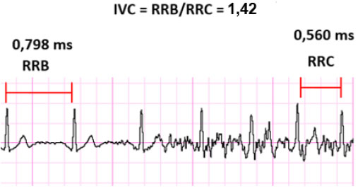 Figure 1. Electrocardiographic tracing of the Cardiac Vagal Index (CVI).
1.2.4 Experimental Procedure - Auriculotherapy
Initially, a disinfection was performed in the auricular pavilions with cotton and 70% alcohol; after, a heating with the fingertips through friction at the apex of the ear (helix), until the characteristic flush was reached, and the bleeding was performed with an specific pen and disposable lancets with a hole in each apex of the ear being drawn from 3 to 5 drops of blood. A 10 cm curved surgical forceps was used to handle the adhesive tape with the seeds.
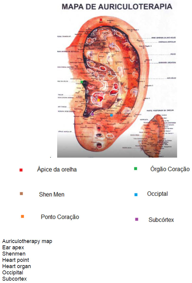
The hypotensive points were stimulated with two mustard seeds arranged in a square of 0.5 x 0.5 cm tape, bilaterally, following the channels and meridians of the ear. The stimulated points were: a) heart - gullet region, with two seeds arranged horizontally; b) heart organ - in the region of the cava shell, two seeds arranged horizontally; c) subcortex – intertragic notch, with two seeds arranged diagonally; d) occipital - end of the lobe together with the scaphoid fossa, with two seeds arranged vertically; e) shenmen - in the region of the triangular fossa, with two seeds arranged diagonally. These atrial points were pressed for five minutes and each point was stimulated for 10 seconds
RESULTS
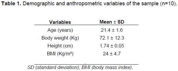
Data on the demographic and anthropometric variables of the 10 individuals evaluated will be presented in Table 1.
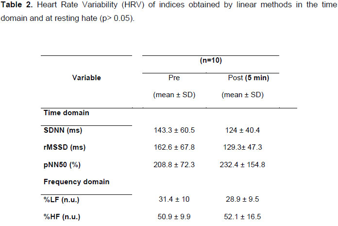
There were no significant differences in relation to variables related to cardiac autonomic control represented by HRV in the domains of time and frequency before and after auriculotherapy (p>0.05), as described in table 2.
The HR measured before and after the evaluation did not show significant differences between the two moments (p> 0.05) (Figure 1).
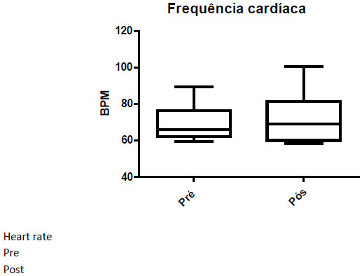 Figure 1. The figure above shows the mean values of pre- and post-auriculotherapy HR.
There was a significant reduction in SBP after acute auriculotherapy of volunteers (p=0.03) (Figure 2).
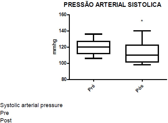 Figure 2. The figure above shows the mean values of the SBP pre- and post-auriculotherapy.
DISCUSSION
The present study aimed to quantify the oscillations that occurred in the ANS modulation, resulting from the auriculotherapy application, verifying the behavior of the sympathetic and parasympathetic branches, using the HRV and T4s tests. Auriculotherapy is based on the understanding that the auricular pavilion has connections with 12 meridians and can restore the balance between blood and vital energy (1, 4, 5, 28). The ear is innervated directly by several pairs of cranial and spinal nerves, dividing into motor and sensory areas, and the vagus nerve is the most important, playing a role in cardiovascular and SNA regulation and affecting mainly the shell region (5,6,16). This study aims to investigate the effectiveness of auriculotherapy in the reduction of HR and BP, by correlating with the physiological changes that occur through ANS. It is well known that auriculotherapy is an alternative treatment, which requires further studies to prove its validity in the scientific field.
The study findings showed a significant reduction in systolic blood pressure variable (SBP) (p<0.05) and a reduction in HRV behavior in 10 individuals, even though there was no significant difference in HRV (p> 0.05). In the HR, there was an increase in this variable, and it was not statistically significant (p=0.59). In a study with patients with some type of pain, hypotensive points such as Shenmen, heart rhythm and contractility zone were stimulated and a significant reduction of HR was observed and 54% reported pain disappearance (29). In a second study, we observed the recovery of athletes who were divided into two groups, one at auriculotherapy group and one control group. The maximum HR was lower than in the control group at 30 and 60 minutes (4). In postmenopausal women, the increase in the sympathetic branch is related to sleep disturbance, and a pre- and post-test study was performed, measuring the HRV before auriculotherapy in 4 weeks of treatment with the points pressed every night. Cardiac discomfort decreased significantly (p<0.05) after 4 weeks (12).
In the literature, there is several evidence linking auriculotherapy to HRV. A study of 12 active patients who had undergone a load test (10 RM), under cardiac monitoring, with mean RR interval differences being significant (p <0.001), and there was a decrease in HRV on exercise, even when performed in a short time (30). In another study with 60 women, atrial points of the liver and spleen were stimulated for 5 days, being an 8-week treatment. Resting HR was observed at baseline and 9 weeks after treatment, with no significant difference between groups, but the sympathetic branch reduced after the fifth week of treatment (9). In a third study, the right thenar muscle was stimulated, there was a significant increase in sympathetic activity, post-stimulation. When the ear was stimulated, there was a significant increase in the parasympathetic branch (p<0.05), indicating that auriculotherapy induces an increase in the HRV high frequency component (21).
There are some variables such as cardiac output, vascular resistance and ejection fraction that may have been determinant for HRV reduction and SBP reduction in the present study, since BP is directly proportional to cardiac output and vascular resistance and, in turn, cardiac output is directly related to HR and ejection fraction, thus there is a hypothesis of other variables that may have influenced the results obtained, since we only analyzed HR and BP. The baroreceptor reflex is a system of BP control, triggered by stretch receptors, which are found in the wall of the systemic arteries, being related to vascular resistance, stimulated above 100 mmHg, altering BP. Studies have shown that auriculotherapy stimulates the baroreceptor system to act on the vagus nerve, increasing parasympathetic activity and modulating CNS and ANS (6, 11, 16, 17). In the HR variable, no significant difference was found, most probably based on two study limitations, which were the sample size of 10 volunteers and also due to the acute application of auriculotherapy, since in some studies the HR reduction was only observed after a considerable time of application, and some evidences showed 30 and 60 minutes auricleotherapy applications or even several weeks of stimulation at a given point. In the frequency domain of the 4-second exercise test, it is observed that SDNN, representing the sympathetic and parasympathetic activity, reduced on auricleotherapy, since rMSSD and pNN50 that represent only the parasympathetic activity, obtained opposite results in auriculotherapy, and rMSSD reduced and pNN50 increased, seen as another factor that may have determined the increase in resting HR. In the frequency domain, sympathetic activity decreased and parasympathetic increased, but not significantly, with equality in sympatho-vagal balance on heart rate. Further studies are needed to determine the contribution of auriculotherapy to cardiovascular treatments.
CONCLUSION
Therefore, our findings suggest a possible contribution of the technique in the reduction of SBP, also demonstrating that there was a behavior of reduction of the HR, being important to verify a longer time of stimulation of the auricular points, in order to analyze its effect on the sympathetic and parasympathetic branches. Therefore, further studies are needed to determine the contribution of auriculotherapy to cardiovascular treatments.
REFERENCES
1. Hou PW, Hsu HC, Lin YW, Tang NY, Cheng CY, Hsieh CL. The History, Mechanism, and Clinical Application of Auricular Therapy in Traditional Chinese Medicine. Evid Based Complement Alternat Med; 2015:495684.
2. Arai YC, Sakakima Y, Kawanishi J, Nishihara M, Ito A, Tawada Y, et al. Auricular acupuncture at the "shenmen" and "point zero" points induced parasympathetic activation. Evid Based Complement Alternat Med; 2013:945063.
3. He W, Rong PJ, Li L, Ben H, Zhu B, Litscher G. Auricular Acupuncture May Suppress Epileptic Seizures via Activating the Parasympathetic Nervous System: A Hypothesis Based on Innovative Methods. Evid Based Complement Alternat Med; 2012:615476.
4. Lin ZP, Chen YH, Fan C, Wu HJ, Lan LW, Lin JG. Effects of auricular acupuncture on heart rate, oxygen consumption and blood lactic acid for elite basketball athletes. Am J Chin Med; 39(6):1131-8.
5. Kim JH, Jung HJ, Kim TH, Lee S, Kim JE, Kang KW, et al. Auricular acupuncture for prehypertension and stage 1 hypertension: study protocol for a pilot multicentre randomised controlled trial. Trials Sep 22; 14:303.
6. Yeh ML, Chang YC, Huang YY, Lee TY. A randomized controlled trial of auricular acupressure in heart rate variability and quality of life for hypertension. Complement Ther Med Apr; 23(2):200-9.
7. Gerhard I, Postneek F. Auricular acupuncture in the treatment of female infertility. Gynecol Endocrinol1992 Sep; 6(3):171-81.
8. Suen LK, Wong TK, Leung AW. Is there a place for auricular therapy in the realm of nursing? Complement Ther Nurs Midwifery2001 Aug;7(3):132-9.
9. Chien LW, Chen FC, Hu HY, Liu CF. Correlation of electrical conductance in meridian and autonomic nervous activity after auricular acupressure in middle-aged women. J Altern Complement Med Aug; 20(8):635-41.
10. Oyola-Santiago T, Knopf R, Robin T, Harvey K. Provision of auricular acupuncture and acupressure in a university setting. J Am Coll Health; 61(7):432-4.
11. Gao XY, Liu K, Zhu B, Litscher G. Sino-European transcontinental basic and clinical high-tech acupuncture studies-part 1: auricular acupuncture increases heart rate variability in anesthetized rats. Evid Based Complement Alternat Med; 2012:817378.
12. Kung YY, Yang CC, Chiu JH, Kuo TB. The relationship of subjective sleep quality and cardiac autonomic nervous system in postmenopausal women with insomnia under auricular acupressure. Menopause Jun; 18(6):638-45.
13. Shu Q, Wang H, Litscher D, Wu S, Chen L, Gaischek I, et al. Acupuncture and Moxibustion have Different Effects on Fatigue by Regulating the Autonomic Nervous System: A Pilot Controlled Clinical Trial. Sci Rep Nov 25; 6:37846.
14. He W, Wang X, Shi H, Shang H, Li L, Jing X, et al. Auricular acupuncture and vagal regulation. Evid Based Complement Alternat Med; 2012:786839.
15. Rong PJ, Zhao JJ, Li YQ, Litscher D, Li SY, Gaischek I, et al. Auricular acupuncture and biomedical research: A promising Sino-Austrian research cooperation. Chin J Integr Med Dec; 21(12):887-94.
16. Gao XY, Li YH, Liu K, Rong PJ, Ben H, Li L, et al. Acupuncture-like stimulation at auricular point Heart evokes cardiovascular inhibition via activating the cardiac-related neurons in the nucleus tractus solitarius. Brain Res Jun 23;1397:19-27.
17. Vanderlei LCM, Pastre CM, Hoshi RA, Carvalho TD, Godoy MF. Noções básicas de variabilidade da frequência cardíaca e sua aplicabilidade clínica. Rev Bras Cir Cardiovasc 2009;24(2):205-17.
18. Rabischong P, Terral C. Scientific Basis of Auriculotherapy: State of the Art. Med Acupunct Apr 1; 26(2):84-96.
19. Ishibashi S. The effect of auricular electroacupuncture on the neuronal activity of the thalamic and hypothalamic neurons of the rat. Acupunct Electrother Res1986;11(1):15-23.
20. Colbert AP, Spaulding KP, A AC, Cutro JA. Clinical utility of electrodermal activity at acupuncture points: a narrative review. Acupunct Med Dec ;29(4):270-5.
21. Haker E, Egekvist H, Bjerring P. Effect of sensory stimulation (acupuncture) on sympathetic and parasympathetic activities in healthy subjects. J Auton Nerv Syst2000 Feb 14;79(1):52-9.
22. de Almeida MB, Ricardo DR, de Araújo CGS. Validação do teste de exercício de 4 segundos em posição ortostática. Arquivos Brasileiros de Cardiologia 2004;83(2):155.
23. Araújo CGS, Ricardo DR, Almeida MB. Fidedignidade intra e interdias do teste de exercício de quatro segundos. Revista Brasileira de Medicina do Esporte 2003;9(5):293-8.
24. Lazzoli JK, Castro CLB, Nobrega ACL, Araújo CGS. Acurácia de critérios para vagotonia no eletrocardiograma de repouso de 12 derivações: uma análise com curvas ROC. Rev Bras Med Esporte 2002; 8:50-8.
25. de Castro CLB, da Nobrega ACL, de Araújo CGS. Testes Autonômicos Cardiovasculares. Uma Revisão Crítica. Parte 11. Arq Bras Cardiol 1992; 59(2):152.
26. Araújo CGS, de Castro CLB, Franca JF, Ramos PS. Teste de Exercício de 4 Segundos: Valores de Referência dos 18 aos 81 Anos de Idade. Arq Bras Cardiol; 104(5):366-74.
27. de Paiva VC, Santana KR, Silva BM, Ramos PS, Lovisi JCM, Araújo CGS, Ricardo DR et al. Comparação entre métodos de avaliação da modulação vagal cardíaca. Arquivos Brasileiros de Cardiologia 97 (6):493-501.
28. Gao XY, Wang L, Gaischek I, Michenthaler Y, Zhu B, Litscher G. Brain-modulated effects of auricular acupressure on the regulation of autonomic function in healthy volunteers. Evid Based Complement Alternat Med 2012:714391.
29. Gaponjuk PJ, Sherkovina TJ, Leonova M. Clinical effectiveness of auricular acupuncture treatment of patients with hypertensive disease. Acupuncture in Medicine 1993; 11(1):29-31.
30. da Paschoa DC, Coutinho JFS, Almeida MB. Análise da variabilidade da frequência cardíaca no exercício de força. Revista da SOCERJ, 2006.