INTRODUCTION
Health Care-Related Infections (HCRI) refer to those acquired during health care delivery, after patient admission, during hospitalization or after discharge1. They represent an important public health problem, both in developed and developing countries, as they increase morbidity and mortality rates, extend hospitalization and raise hospital costs1,2.
Despite important advances in infection control, studies have demonstrated the emergence of the problem globally, with a marked increase in the frequency and severity of cases of HCRI3,4. In the United States, more than 70% of the bacteria isolated in hospitals are resistant to at least one antimicrobial commonly used in the treatment of infection5. The acquisition of resistant microorganisms usually occurs due to the non-adherence of the professionals to biosafety measures, the work overload, sometimes the reduced number of human resources and the direct contact of the patient with environment or contaminated material4,5.
Currently, it is believed that a considerable proportion of HCRI can be avoided, and hand hygiene is still very important in this context because it is a measure of unquestionable effectiveness and low cost, since the microorganisms most associated with the occurrence of such infections belong to the transient microbiota (acquired through contacts established with persons colonized or infected and/or with contaminated objects). These microorganisms can be eliminated through hand hygiene, which, when not performed or performed inadequately, is a basic premise for the occurrence of cross-transmission6,7.
Hand hygiene along with correct technique should be part of the routine of health professionals. Well-performed studies have shown the importance of this practice in the reduction of HCRI rates and the majority of specialists in infection control affirm that this procedure is the simplest and most effective means of preventing the transmission of microorganisms in the care environment8,9.
Hand-washing with soap and water eliminates transient microorganisms and reduces residents and, most of the time, disrupts the disease transmission chain. As a way to prevent infections the action of hand hygiene is shown as a great measure, but it is important to remember that the soaps and antiseptics used during this process can be an important vehicle of contamination, which contamination can occur during the process (intrinsic contamination) or during its use (extrinsic contamination)10,11.
Considering the above mentioned, this study aimed to evaluate the extrinsic contamination by pathogenic bacteria of liquid soaps used during the handwashing process of health professionals in a philanthropic hospital.
METHODS
The design adopted in this study was cross-sectional descriptive, where we evaluated the presence of extrinsic contamination of liquid soaps used in a philanthropic hospital.
The samples were collected in the months of August and September 2016, of plastic soap holders, fixed to the wall, with refill for replenishment and soap dispenser. With sterile swab with Stuart medium (Labor® Swab) the sample was collected from the push and 1 ml of the liquid soap was collected in the test tube. After the collection, the samples were sent to the microbiology laboratory of the Faculty of Medical Sciences and Health of Juiz de Fora.
The samples were inoculated in BHI broth (Mbiolog diagnostics Ltda.®), incubated for 48 hours in an aerobiose oven (Fanem® 502 São Paulo - Brazil), at 35ºC ± 1. After this incubation period, they were spiked in Blood Agar 5 (RenyLab®, Química e Farmacêutica) and incubated for 24-48 hours in aerobiosis at 35º ± 1ºC and biochemical tests were performed to identify each species12,13. For the identification of Gram-negative rods, OF, IAL, citrate, arginine, ornithine, lysine, motility, oxidase and resistance to polymyxin B were used12,13.
The bacteria identified underwent antimicrobial susceptibility test (AST) by the Disk Diffusion Agar method, according to the standardization of the CLSI 2016 - Clinical Laboratory Standards Institute14.
For the Enterobacteriaceae family, the following antimicrobials were tested: amoxicillin + clavulanate, ceftazidime, ceftriaxone, cefepime, aztreonam, ertapenem, meropenem, imipenem, gentamicin, amikacin, sulfamethoxazole + trimethoprim, ciprofloxacin and levofloxacin.
For Acinetobacter spp., ceftazidime, ceftriaxone, cefepime, meropenem, imipenem, gentamicin, amikacin, sulfamethoxazole + trimetoprin, levofloxacin, ciprofloxacin, ampicillin + sulbactam and tetracycline were tested. And for pseudomonas spp., piperacillin + tazobactam, caftazidime, cefepime, aztreonam, gentamycin, imipenem, meropenem, amikacin, levofloxacin, ciprofloxacin were tested.
RESULTS
32 soap dispensers were analyzed, totalizing 64 samples, 32 soap dispenser pushbuttons and 32 liquid soaps used in different sectors of the hospital, being divided into Intensive Care Unit 1 - ICU1 (14 samples), Neonatal Intensive Care Unit - NICU (10 samples), Intensive Care Unit 2 - ICU2 (12 samples), Intensive Care Unit Reception Room 1 - ICURR1 (4 samples), Surgical Center - SC (4 samples), Normal Delivery Center - NDC (4 samples), Inpatient Unit 5 - INPU5 (4 samples), Inpatient Unit 6 -INPU6 (8 samples) and in the Canteen - CAN (4 samples).
Of the 64 samples collected, in 25 (39.09%) there was growth of bacteria, 14 (21.87%) in the dispensing push buttons and 11 (17.19%) in soaps. The percentage of microbial growth according to the sampled site is described in figure 1. In 4.68% of the samples there was growth of more than one bacterial species.
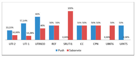 Figure 1. Percentage of microbial growth in dispensing push buttons and liquid soaps, distributed by hospital sectors.
In relation to the microorganisms found, of the 25 positive samples of dispensing push buttons and soaps, 28 strains were identified, being 12 strains (42.85%) of Acinetobacter spp, 4 (14,28%) of Enterobacter cloacae, 3 10.72%) of Klebsiella oxytoca, 3 (10.72%) of Pantoea spp., 3 (10.72%) of Pseudomonas spp., 2 (7.14%) Escherichia coli, and 1 (3.57 %) of Klebsiella pneumoniae as described in figure 2.
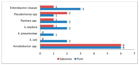 Figure 2. Distribution of the number of bacterial species found in the dispensing push buttons and soaps of a philanthropic hospital.
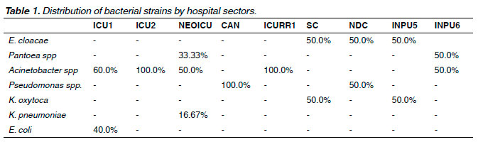
In the ICU1, 60.0% of the positive samples were Acinetobacter spp. and 40.0% E. coli. In NICU the prevalence was 50.0% of Acinetobacter spp., 33.33% of Pantoea spp. and 16.67% of K. pneumoniae. In ICU2 the growth was 100.0% of Acinetobacter spp., and in the NDC it was 50.0% of Pseudomonas spp. and 50.0% E. cloacae. In ICURR1 it was 100.0% of Acinetobacter spp. and in the CAN it was 100.0% of Pseudomonas spp. In INPU5 it was 50.0% of K. oxytoca and 50.0% of E. cloacae, and in INPU6 it was 50.0% of Acinetobacter spp. and 50% of Pantoa spp. In SC it was 50.0% of E. cloacae and 50.0% of K. oxytoca, according to table 1.
The Antimicrobial Sensitivity Test (TSA) was performed to evaluate the resistance profile of the bacteria found. The antimicrobials tested were in accordance with the CLSI 2016 standardization, and the observed resistance profile is reported in table 2.
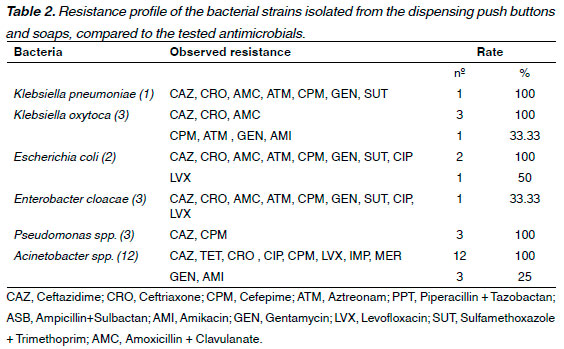
By means of phenotypic tests, E. coli strains (2), K. oxytoca (1), K. pneumoniae (1) and E. cloacae (1) were found to be extended-spectrum beta-lactamase (ESBL) producers, and the Pseudomonas sp. strains (3) were AmpC beta-lactamase producing agents.
DISCUSSION
Most environments are susceptible to contamination by microorganisms directly related to the local hygienic situation. Consequently, objects of routine use and with inadequate hygiene may become foci of contamination and infection in susceptible hosts. The survival of microorganisms in the environment varies on different types of surfaces, varying from a few minutes to months. The longer a microorganism persists viable on a surface, the longer it remains as a source of transmission, thus increasing the chance of transfer to a host15.
A central issue in the prevention and control of infections is the importance of handwashing as the primary effort of health professionals to prevent and spread infectious diseases in the clinical setting16,17. However, liquid soap can become contaminated with bacteria and represent a recognized health risk in health care settings16,18. In particular, reusable soap dispensers are prone to bacterial contamination and several outbreaks associated with the use of contaminated soap in health care settings have been reported10,16,18-21.
Manufacturers provide instructions for handwashing, but typically do not provide instructions for recharging dispensers. Such instructions would be beneficial because liquid soaps are purchased in gallons to recharge the dispensers18. In addition, when reusable pumps are supplied and transferred into several bottles, cross-contamination is a concern. Regular observation of expiration dates is important because active or preservatives agents may degrade over time, which may reduce the effectiveness of soap against microorganisms18, 22.
Open dispensing systems, according to the practice used in the institution of the present study, and the manipulation of liquid soaps in the hospital environment are known risk factors for the microbial contamination of these products10. Some studies make recommendations to prevent soap from becoming a source of infection, such as buying disposable dispensers, using soap bottles with smaller volumes, limiting the use of liquid soap, providing individual soap to patients, and increasing disinfection with alcohol gel10,23.
Although facilities may have documented cleaning protocols for liquid soap dispensers, noncompliance may be a problem that leads to contamination. Several studies have investigated the microbial contamination of liquid soaps in hospital and community settings. A number of opportunistic bacterial pathogens were recovered from liquid soaps, including Klebsiella pneumoniae, Serratia marcescens, methicillin-resistant Staphylococcus aureus (MRSA), Pseudomonas sp. and Enterobacter spp.16,20.
In the present study, it was found that 39.06% of the samples were contaminated. Twelve strains of Acinetobacter spp., the main pathogen identified, were found. Acinetobacter spp. has been responsible for causing different types of infections, such as pneumonia, septicemia, urinary infections and meningitis, especially in immunocompromised patients, and is considered an opportunistic pathogen of great importance in HCRI24. During the last decade, the treatment of these infections has become critical due to the emergence of multiresistant strains whose dissemination has been associated with the contamination of hospital equipment (respirators, air conditioning, imaging equipment, etc.) and/or through contaminated hands of the care team24,25.
Pseudomonas spp. is a ubiquitous microorganism found easily in any hospital setting and is an important agent of HCRI frequently identified as a causal agent of infection due to its ability to survive in places of high humidity such as sinks and breathing apparatus as ventilation masks26. Almost all hospital equipment and materials, mainly with liquid components, can serve as reservoirs for Pseudomonas spp. including respiratory ventilation equipment, intravenous therapy and even some germicides such as liquid soap and disinfectants10,26. Intrinsic resistance to multiple antimicrobials contributes to the opportunistic nature of this pathogen26,27. It was found in this study that 100% (3) of the strains of Pseudomonas sp. were AmpC beta-lactamase producing agents, an enzyme that confers resistance to cephalosporins and penicillins.
It was observed the growth of Enterobacteriaceae strains, 5 were ESBL producers, being E. coli (2), K. oxytoca (1), K. pneumoniae (1) and E. cloacae (1). The production of ESBL by enterobacteria has an important impact on the morbidity and mortality rates as well as the costs of hospital and community treatment28-30. The production of ESBL is an important mechanism of resistance in enterobacteria29. The treatment of infections caused by ESBL-producing strains presents a substantial challenge to antimicrobial therapy since ESBLs are able to hydrolyze penicillins, cephalosporins of all generations and monobacchaeam, minimizing the therapeutic options29-33. In addition, the continuous and often inadequate use of antimicrobial agents may induce the selection of multiresistant strains29,34.
In this study, we observed the growth of Pantoea spp. in dispensers and soap, with prevalence of 3 strains, which did not present resistance to the tested antimicrobials. Pantoea spp. is an important plant pathogen that can cause disease in rare cases and has already been found in wounds, blood and urine as an opportunistic pathogen and has been responsible for several national sepsis outbreaks in the US12,35. In the mid-1960s Pantoea agglomerans were identified in HCRI, being the most frequent species associated with human infections36,37. Hospital outbreaks due to contamination of anesthetic propofol, blood products, parenteral nutrition, and transfer tubes used for intravenous hydration have been reported35-38.
P. agglomeransis a causative agent of infection in children and the elderly and can cause bacteremia, often in association with more conventional pathogens, and is also responsible for outbreaks in NEOICU35,36,38,39. In recent years a remarkable increase in HCRI has been reported, especially in the NEOICU, intensive care units and oncology. Baseline diseases, low birth weight, immunocompromised immune system, chemotherapy, and the use of invasive devices may be predisposing factors in infections caused by unusual microorganisms, including P. agglomerans36,37.
Based on these considerations, this study evaluated the extrinsic contamination of liquid soaps used in a philanthropic hospital, since, in our view, studies of this nature help prevent infection and, consequently, reduce their rates and costs. Thus, they have benefits for the institution and for the patient who will not have his health condition compromised nor his prolonged stay in the hospital due to an HCRI. Although it cannot be stated herein that they have influenced infection rates, since the causal relationship has not been established, some bacteria are known agents of HCRI. The confirmed presence of pathogenic bacteria in soaps of hospital use awakens to the necessity of more rigid measures of disinfection of the hospital area.
CONCLUSION
There were extrinsic contamination of the liquid soaps used for handwashing in different sectors of the hospital by pathogenic bacteria such as Acinetobacter spp, Enterobacter cloacae, Klebsiella oxytoca, Pantoea spp., Pseudomonas spp., Escherichia coli andKlebsiella pneumoniae.
The isolated bacterial strains showed high bacterial resistance, and resistance mechanisms such as ESBL and AmpC were observed.
Thus, considering all the possibilities of microorganisms isolated in this study to demonstrate resistance to antimicrobials, favoring infection in immunocompromised patients, it is of the utmost importance that the participants of the hospital infection commission also include among the materials inspected during their inspections the products used mainly for the prevention of infections, such as soaps and antiseptics.
REFERENCES
1. Albuquerquer AM, Souza APM, Torquato IMB, Trigueiro JVS, Ferreira JA, Ramalho MAN. Infecção Cruzada no Centro de Terapia Intensiva á Luz da Literatura. Rev. Ciênc. Saúde Nova Esperança 2013; 11 (1):78-87.
2. Oliveira AC, Silva MDM, Garbaccio JL. Vestuário de profissionais de saúde como potenciais reservatórios de microrganismos: Uma revisão integrativa. Texto Contexto Enferm 2012; 21 (3): 684-91.
3. Oliveira AC, Damasceno QS. O papel do ambiente hospitalar na disseminação de bactérias resistentes. Rev Epidemiol Control Infect 2012; 2 (1):28-31.
4. Correal JCD, Marques EA, Guilherme WL, Leão RS, Damasco PV. Infecções por Staphylococcus aureus: mudança do perfil epidemiólogico no Hospital Universitário Pedro Ernesto. Rev HUPE 2013; 12 (3):31-46.
5. Oliveira AC, Cardoso CS, Mascarenhas D. Precauções de contato em unidade de terapia intensiva: fatores facilitadores e dificultadores para adesão dos profissionais. Rev Esc Enferm USP 2010; 44 (1): 161-5.
6. Martinez J, Roseira CE, Passos IPBD, Figueiredo RM. Higienização das mãos: conhecimento dos estudantes. Cienc Cuid Saude 2014; 13 (3): 455-63.
7. Oliveira AC. Infecções hospitalares: repensando a importância da higienização das mãos no contexto de multirresistência. Rev Min Enf 2003; 7 (2): 140-4.
8. Brasil. Ministério da Saúde. Programa Nacional de Prevenção e Controle de Infecções Relacionadas à Assistência à Saúde. Brasília, 2013.
9. Mota EC, Barbosa DA, Silveira BRM, Rabelo TA, Silva NM, Silva PLN, et al.,. Higienização das mãos: uma avaliação da adesão e da prática dos profissionais de saúde no controle das infecções hospitalares. Rev Epidemiol Control Infect 2014; 4 (1): 12-7.
10. Caetano JA, Lima MA, Miranda MDC, Serufo JC, Pontes PRL. Identificação de contaminação bacteriana no sabão líquido de uso hospitalar. Rev Esc Enferm USP 2011; 45 (1): 153-60.
11. Moreira ACA, Carvalho JLM. Ocorrência de Klebsiella pneumoniae e outros coliformes em sabão neutro líquido utilizado em um berçário de hospital. Rev Ciênc Med Biol 2006; 5 (3): 245-52.
12. Koneman EW. Diagnóstico Microbiológico: texto e atlas. 6 ed. Rio de Janeiro: Guanabara Koogan; 2008.
13. Oplustil CP, Zoccoli CM, Tobouti NR. Procedimentos Básicos em Microbiologia Clínica. 3 rd ed. Rio de Janeiro: Sarvier; 2010.
14. Clinical Laboratory Standards Institute. Methods for dilution antimicrobial susceptibility test for bacteria that grow aerobically 2016.
15. Alves JLB, Costa RM, Braoios A. Teclados de computadores como reservatórios de microrganismos patogênicos. J Health Sci Inst. 2014; 32 (1): 7-11.
16. Momeni SS, Tomlin N, Ruby JD. Isolation of Raoultella planticola from refillable antimicrobial liquid soap dispensers in a dental setting. JADA 2015; 146 (4): 241-5.
17. Souza LM, Ramos MF, Becker ESS, Meirelles LCS, Monteiro SAO. Adesão dos profissionais de terapia intensiva aos cinco momentos da higienização das mãos. Rev Gaúcha Enferm. 2015; 36 (4): 21-8.
18. Zapka CA, Campbell EJ, Maxwell SL, et al. Bacterial hand contamination and transfer after use of contaminated bulk-soap-refillable dispensers. Appl Environ Microbiol. 2011; 77 (9): 2898-904.
19. Archibald LK, Corl A, Shah B, Schulte M, Arduino MJ, Aguero S, Fisher DJ et al. Serratia marcescens outbreak associated with extrinsic contamination of 1% chlorxylenol soap. Infect. Control Hosp. Epidemiol. 1997; 18: 704–9.
20. Buffet-Bataillon S, Rabier V, Bétrémieux P, Beuchée A, Bauer M, Pladys P et al. Outbreak of Serratia marcescens in a neonatal intensive care unit: contaminated unmedicated liquid soap and risk factors. J. Hosp. Infect. 2009; 72: 17–22.
21. Weber D J, Rutala WA, Sickbert-Bennett EE. Outbreaks associated with contaminated antiseptics and disinfectants. Antimicrob. Agents Chemother. 2007; 51: 4217–24.
22. Serufo JC. Avaliação da dinâmica de contaminação extrínseca de sabonetes líquidos e anti-sépticos no processo de uso em hospitais brasileiros da rede sentinela. Belo Horizonte: Fundação de Desenvolvimento da Pesquisa (FUNDEP) e Agência Nacional de Vigilância Sanitária (ANVISA), 2009.
23. Assadian O, Kramer A, Christiansen B, Exner M, Martiny H, Sorger A et al. Recommendations and requirements for soap and hand rub dispensers in healthcare facilities. GMS Krankenhaushyg Interdiszip.2012; 7 (1): Doc03.
24. Martins AF, Barth AL. Multidrug-resistant Acinetobacter – a challenge for public health. Scientia Medica 2013; 23 (1): 56-62.
25. Giamarellou H, Antoniadou A, Kanellakopoulou K. Acinetobacter baumannii: a universal threat to public health? Int J Antimicrob Agents. 2008; 32 (2):106-19.
26. Blanc DS, Gomes Magalhaes B, Abdelbary M, Prod'hom G, Greub G, Wasserfallen JB, Genoud P, Zanetti G, Senn L, Hand soap contamination by Pseudomonas aeruginosa in a tertiary care hospital: no evidence of impact on patients. J Hosp Infect. 2016; 93 (1): 63-7.
27. Lima ABM, Leão-Vasconcelos LSNO, Costa DM, Vilefort LOR, André MCDPB, Barbosa MA et al. Pseudomonas spp. isolated from the oral cavity of healthcare workers from an oncology hospital in midwestern Brazil. Rev Inst. Med Trop 2015; 57 (6): 513-4.
28. Kaiser TDL, Santiago DD, Mendes EMT, Matos BV. Detecção de betalactamase de espectro estendido em isolados de enterobactérias provenientes de um hospital da região de Santa Teresa-ES. Arq. Ciênc. Saúde 2016; 20 (1): 3-7.
29. Lago A, Fuentefria SR, Fuentefria DB. Enterobactérias produtoras de ESBL em Passo Fundo, Estado do Rio Grande do Sul, Brasil. Rev Soc Bras Med Trop 2010; 43 (4): 430-4.
30. Lenhard-Vidal A, Cardoso RF, Pádua RAF, Siqueira VLD. High prevalence rate of extended-spectrum beta-lactamases (ESBL) among Enterobacteriaceae in a small Brazilian public hospital. Brazilian Journal of Pharmaceutical Sciences 2011; 47 (4): 701-7.
31. Ouedraogo AS , Sanou M , Kissou A , Sanou S , Solaré H , Kaboré F et al. High prevalence of extended-spectrum β-lactamase producing enterobacteriaceae among clinical isolates in Burkina Faso. BMC Infectious Diseases 2016: 16: 326.
32. Singh N, Pattnaik D, Neogi DK, Jena J, Mallick B. Prevalence of ESBL in Escherichia coli Isolates Among ICU Patients in a Tertiary Care Hospital. Journal of Clinical and Diagnostic Research. 2016; 10 (9): 19-22.
33. Enoch DA , Brown F , Sismey AW , Mlangeni DA , Curran MD , Karas JA et al. Epidemiology of extended-spectrum beta-lactamase-producing Enterobacteriaceae in a UK district hospital; an observational study. J Hosp Infect. 2012: 81 (4): 270-7.
34. Wollheim C, Guerra IMF, Conte VD, Sheila P Hoffman SP, Schreiner FJ, Delamare APL et al. Nosocomial and community infections due to class A extended-spectrum b lactamase (esbla)-producing Escherichia coli and Klebsiella spp. in southern Brazil. Braz J Infect Dis 2011; 15 (2): 138-43.
35. Sengupta M, Banerjee S, Das NK, Guchhait P, Misra S. Early Onset Neonatal Septicaemia Caused by Pantoea agglomerans. Journal of Clinical and Diagnostic Research 2016: 10 (5): 1-2.
36. Mardaneh J, Dallal MMS. Isolation, identification and antimicrobial susceptibility of Pantoea (Enterobacter) agglomerans isolated from consumed powdered infant formula milk (PIF) in NICU ward: First report from Iran. Iran J Microbiol 2013; 5 (3): 263-7.
37. Boszczowski I, Nóbrega de Almeida Júnior J, Peixoto de Miranda EJ, Pinheiro Freire M, Guimarães T, Chaves CE, et al. Nosocomial outbreak of Pantoea agglomerans bacteraemia associated with contaminated anticoagulant citrate dextrose solution: new name, old bug?. J Hosp Infect. 2012; 80: 255–8.
38. Mehar V, Yadav D, Sanghvi J, Gupta N, Singh K. Pantoea dispersa: an unusual cause of neonatal sepsis. Braz J Infect Dis. 2013;17(6):726–8.
39. Van Rostenberghe H, Noraida R, Wan Pauzi WI, Habsah H, Zeehaida M, Rosliza AR, et al. The clinical picture of neonatal infection with Pantoea species. Jpn J Infect Dis. 2006; 59: 120–1.