
INTRODUÇÃO: A trombocitopenia aloimune fetal e neonatal (FNAIT) é causada por incompatibilidade de antígenos plaquetários entre mãe e feto, que resulta em respostas imunológicas mediadas por anticorpos. O organismo da mãe produz anticorpos contra antígenos plaquetários fetais específicos. Esses anticorpos atravessam a placenta e ligam-se às plaquetas fetais, removendo-as da circulação através do sistema reticuloendotelial.
OBJETIVO: Analisar, por meio de revisão sistemática, os casos de FNAIT para verificar a classe de antígenos plaquetários mais envolvida com a patologia e compreender as medidas preventivas adotadas durante a gestação.
MÉTODO: Foi realizada uma busca de artigos científicos dos últimos 10 anos. A base de dados utilizada foi o PubMed, sendo os descritores “Thrombocytopenia, Neonatal Alloimmune” e “Clinical Laboratory Techniques” determinados pelo MesH (Medical Subject Headings). Foram aplicados critérios de inclusão com base na data de publicação, espécie e idioma. Os artigos considerados não relevantes e estudos do tipo “Relatos de Caso” foram excluídos.
RESULTADOS: Foram selecionados para fazer parte do escopo desta revisão 7 artigos, que apontaram as incompatibilidades envolvendo o HPA-1a como as principais responsáveis pelos quadros de FNAIT. Com relação aos tratamentos pré-natais oferecidos às gestantes, observou-se uma variabilidade nas estratégias terapêuticas, mas o emprego da imunoglobulina intravenosa associada ou não à corticosteróides têm se mostrado eficaz para a prevenção de hemorragia intracraniana e óbitos de fetos/neonatos.
CONCLUSÃO: Os casos de FNAIT, apesar de raros, são graves e mostraram-se de difícil diagnóstico e manejo clínico. Portanto, há necessidade de desenvolvimento contínuo de novos estudos que auxiliem no diagnóstico e na padronização de métodos preventivos.
Palavras-chave: Trombocitopenia Neonatal Aloimune, Imunização, Hemorragias Intracranianas, Morte Fetal.
INTRODUCTION
Fetal and neonatal alloimmune thrombocytopenia (FNAIT) is the most common cause of thrombocytopenia in neonates and affects between 1/800 and 1/1,000 live births, and the occurrence of severe cases (counting platelets <50,000/mm³) caused by anti-HPA-1a antibodies is approximately ½,500 pregnancies1-3.
FNAIT is caused by an incompatibility of platelet antigens between the mother and the fetus, which results in antibody-mediated immune responses. The mother’s body produces antibodies of the immunoglobulin G (IgG) type against specific fetal platelet antigens that are of paternal origin. These produced antibodies cross the placental barrier and bind to fetal platelets, which will later be removed from the circulation by the reticuloendothelial system resulting in fetal thrombocytopenia1,4.
Alloimmunization occurs for one or more human platelet antigens (HPA). These antigens are located on platelet glycoproteins, and most of which are located on the transmembrane glycoprotein GPIIb/IIIa. It was found that the antibodies involved with FNAIT are related to six types of biallelic antigens (HPA-1, HPA-2, HPA-3, HPA-4, HPA-5, and HPA-15). The different HPA alleles vary according to ethnic groups, with the antibody against HPA-1a being the most prevalent in Caucasians, responsible for 70 to 85% of cases of FNAIT in this population5.
Babies who develop FNAIT have thrombocytopenia, petechiae, and bruises. In addition, about 10% of cases may progress to intracranial hemorrhage (ICH), the most serious complication that can lead to death or neurological disability in approximately 90% of cases6,7.
As there is no routine screening for all pregnant women, there is no way to predict the occurrence of FNAIT today. Therefore, to reduce cases of complications for ICH, the intervention method is the treatment of pregnancies subsequent to a previous pregnancy identified with FNAIT. However, only 13% of cases are diagnosed using this obstetric history as a predictor. In prenatal examinations, preventive measures are adopted for high-risk pregnancies, such as weekly infusion of intravenous immunoglobulin (IVIG) to the mother, associated or not with corticosteroids, and intrauterine platelet transfusion. Initially, a fetal blood sample is taken for platelet count and then, if necessary, intrauterine transfusion of compatible platelets is performed. However, these measures are associated with premature births and abortions in 1 to 2% of the procedures2,7.
Given the severity of FNAIT and the challenge for its diagnosis even in the prenatal period, the aim of this systematic review was to analyze, from a laboratory and therapeutic perspective, the cases of fetal and neonatal alloimmune thrombocytopenia to check the class of platelet antigens most involved in the cases and understand the preventive measures adopted during the gestational period.
METHODS
Research strategies
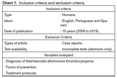
A search for scientific articles from the last 10 years was performed, until August 31, 2019. The database used was PubMed, with the descriptors “Thrombocytopenia, Neonatal Alloimmune” and “Clinical Laboratory Techniques” determined by MesH (Medical Subject Headings). Using the advanced search tool through the Boolean operators “OR” and “AND” 763 articles were found. Then, some inclusion criteria were applied based on the date of publication, species, and language. Articles considered not relevant and “Case Reports” studies were excluded (Chart 1).
RESULTS
763 scientific articles were identified involving alloimmune neonatal/fetal thrombocytopenia and clinical laboratory techniques. However, after applying the previously established inclusion criteria, 227 studies remained for reading the titles. Excluding those considered not relevant, 79 articles were chosen to read the abstracts and “Case Report” studies were excluded. Finally, 7 articles were selected to be part of the scope of this systematic review. Figure 1 shows the flowchart of the article selection process.
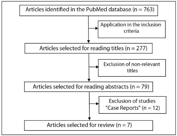
Figure 1. Flowchart of the article selection process.
In general, the studies analyzed involved different approaches, such as: I) retrospective analysis of cases of FNAIT or alloimmunization by HPA-1a; II) follow-up studies of women who received prenatal treatments in their pregnancies, as they already had children with ICH; and III) comparisons between previous pregnancies and subsequent pregnancies of the same women who had children with FNAIT (Table 1).
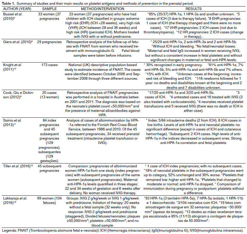
As has already been described in the literature, all studies in the scope of this systematic review pointed out the incompatibilities involving the HPA-1a antigen as the main responsible for the FNAIT presentations, with prevalence ranging from 85 to 95% of cases. The authors demonstrated the importance of a more in-depth assessment of FNAIT or ICH history in previous pregnancies in order to adopt preventive measures for subsequent pregnancies. Regarding the prenatal treatments offered to pregnant women, it was possible to observe variability in the therapeutic strategies adopted by hospitals or therapy centers, but, despite this heterogeneity, the use of IVIG associated or not with corticosteroids has been shown to be effective for the prevention of ICH and deaths of fetuses or neonates.
DISCUSSION
The results showed the importance of preventive methods in the prenatal period for pregnant women who had a history of ICH or FNAIT in previous pregnancies, despite the therapeutic regimens involving IVIG and corticosteroids vary between studies.
Efficacy of therapies in the prenatal period for preventing ICH or deaths in fetuses/neonates
Studies have shown the importance of therapies with intravenous immunoglobulin, associated or not with corticosteroids and intrauterine platelet transfusions, to minimize the occurrence of hemorrhagic complications, such as ICH, which can result in deaths of fetuses/neonates.
This can be demonstrated in the study by Cook et al. (2012)6, in which 16 women were treated with IVIG 1 g/kg/week, and three of whom received an association with corticosteroids (prednisolone), four neonates received platelet transfusions and five received IVIG after birth and there was no ICH or death in these cases. A similar approach was performed in the study by Giers et al. (2010)9 where pregnant women were also treated with IVIG 1 g/kg/week, but, before delivery, the fetuses received intrauterine platelet transfusion: there were no hemorrhagic complications and no deaths, despite not showing a consistent increase in blood count fetal platelets.
In the study by Lakkaraja et al. (2016)13, whose aim was to assess whether the empirical therapy followed by scheduling would be effective and would allow omission of fetal blood collection, two treatment groups were created: I) IVIG 2 g/kg/week divided into two infusions in the week and II) IVIG 1 g/kg/week with 0.5 mg/kg/day prednisone, as empirical therapies initiated without fetal sampling, being performed only in the 32nd week of pregnancy. In this protocol, those who did not respond to therapy in fetal sampling, had an increase to IVIG 2 g/kg/week and 0.5 mg/kg/day prednisone (staggered therapy). Therefore, after fetal blood collection, 19/104 fetuses had platelets <50,000/mm³ and 13 received staggered therapy: 11 of these were born with platelets > 50,000/mm³ and 3/104 neonates suffered postnatal ICH. Then, these authors concluded that the increase in fetal platelet count occurred in almost all cases and that these two forms of initial treatment followed by escalation are reasonably safe and effective.
Bussel et al. (2010)8, who studied the prevention of ICH through prenatal therapy with pregnant women from two consecutive studies, classified women into risk groups and different forms of therapy adopted, with adjustments in dosages being made: for example, a pregnant woman from the high-risk group was treated with IVIG 1 g/kg/week and prednisone 1 mg/kg/day but, after losing the baby, the next pregnant women in this group were treated with IVIG 2 g/kg/week divided into two doses. The pregnancies in the first study in the very high-risk group received IVIG 1 g/kg/week and in the second IVIG study 1 g/kg/week or 2 g/kg/week. Finally, the pregnancies of the high-risk group in the first study received IVIG 1 g/kg/week and prednisone 1 mg/kg/day or IVIG 1 g/kg/week only and, in the second study, IVIG 1 g/kg/week or IVIG 2 g/kg/week.
Through the analysis of these studies taken as an example, it is noticed that different methods of prevention of hemorrhagic complications are adopted that can evolve to fetal/neonate losses, with variations in intravenous immunoglobulin dosages as well as the corticosteroid used (prednisone or prednisolone) and its dosages.
Laboratory techniques for FNAIT diagnosis
Regarding the techniques used by the authors to identify and quantify antibodies against HPA, Giers et al. (2010)9, Sainio et al. (2013) 11 and Tiller et al. (2016)12 used the immobilization test for platelet antigens by monoclonal antibodies (MAIPA).
MAIPA is an assay that uses a donor platelet panel with known genotyping and specific monoclonal antibodies against the glycoproteins that contain the target antigens, that is, when using a monoclonal antibody against glycoprotein IIbIIIa, the presence of antibodies against specific antigens of this complex is assessed, such as HPA-1 and HPA-3. As it is a totally “in-house” technique, it requires good standardization through the use of internal and external controls. In addition, it is a technically demanding test, requiring up to 8 hours to be performed and requires a high level of technical knowledge to interpret its results. For this reason, other methods for detecting these antibodies have been developed14,15.
In addition to this, other techniques are also used, such as platelet immunofluorescence tests (PIFT) and enzyme-linked immunosorbent assay (ELISA), based on the use of purified platelet membrane glycoproteins. However, as MAIPA is the most sensitive and a specific technique for detecting alloantibodies against HPA, it has been considered the gold standard method for the diagnosis of FNAIT16,17.
Fetal sampling and its complications
Authors of two studies pointed out complications associated with the procedure for collecting fetal blood samples. For example, Bussel et al. (2010)8 reported that this may have been the cause of premature delivery at 24 weeks of gestation. In addition, after fetal blood collection, there was an occurrence of fetal tachycardia and eight occurrences of bradycardia, and in three of these cases an emergency cesarean section was necessary, in which one was due to hemorrhage at the needle perforation site. In the study by Lakkaraja et al. (2016)13, which even aimed to assess whether fetal blood collection can be omitted through empirical therapy, of eighty women who underwent the procedure, 12.5%10 suffered complications, such as premature birth or rupture of membranes in six cases and, in the remaining four cases, a cesarean section was performed due to fetal bradycardia. Finally, these authors concluded that the initial treatment protocols followed by empirical scheduling of therapy is reasonably safe, effective in increasing fetal platelets and allows the omission of fetal blood collection.
CONCLUSION
The present review shows that the majority of cases of mother/fetus incompatibility of platelet antigens involved the anti-HPA-1a class, as it has been described in the literature. In addition, the study demonstrates a variability in the therapeutic strategies adopted for prevention in high-risk pregnancies, but, despite this, the use of IVIG associated or not with corticosteroids proved to be effective for the prevention of ICH and deaths of fetuses or neonates.
FNAIT cases, although rare, are serious and have proved to be difficult to diagnose and manage clinically. Therefore, there is a need for continuous development of new studies that assist in the diagnosis of the disease and the standardization of effective preventive methods during the prenatal period.
REFERENCES
1. Kamphuis MM, Oepkes D. Fetal and neonatal alloimmune thrombocytopenia: prenatal Interventions. Prenat Diagn 2011; 31:712-719.
2. Kjær M, Bertrand G, Bakchoul T, Massey E, Baker JM, Lieberman L, et al. Maternal HPA-1a antibody level and its role in predicting the severity of fetal/neonatal alloimmune thrombocytopenia: a systematic review. Vox Sanguinis 2019; 114:79-94.
3. Poles A, Lucas G, Green F, Walser P, Davey S, Ridgwell K, et al. Neonatal alloimmune thrombocytopenia due to a new alloantigen Bl(a) defined by an Asp458Gly substitution in GPIIIa. Transfusion 2019; 59:396-404.
4. Refsum E, Meinke S, Gryfelt G, Wikman A, Höglund P. Adding to the complexity of fetal and neonatal alloimmune thrombocytopenia: reduced fibrinogen binding in the presence of anti-HPA-1a antibody and hypo-responsive neonatal platelets. Thrombosis Research 2018; 162:69-76.
5. Bonstein L, Haddad N. Taking a wider view on fetal/neonatal alloimmune thrombocytopenia. Thrombosis Research 2017; S100-S102.
6. Cook TJ, Qiu CC, Dickinson JE. A review of the contemporary management of fetal and neonatal alloimmune thrombocytopenia in an australian tertiary obstetric hospital. Australian and New Zealand Journal of Obstetrics and Gynaecology 2012; 52:321-326.
7. Wienzek-Lischka S, Krautwurst A, Fröhner V, Hackstein H, Gattenlöhner S, Bräuninger A, et al. Noninvasive fetal genotyping of human platelet antigen-1a using targeted massively parallel sequencing. Transfusion 2015; 55;1538-1544.
8. Bussel JB, Berkowitz RL, Hung C, Kolb EA, Wissert M, Primiani A, et al. Intracranial hemorrhage in alloimmune thrombocytopenia: stratified management to prevent recurrence in the subsequent affected fetus. Am J Obstet Gynecol 2010; 203:135.e1-14.
9. Giers G, Wenzel F, Stockschläder M, Riethmacher R, Lorenz H, Tutschek B. Fetal alloimmune thrombocytopenia and maternal intravenous immunoglobulin infusion. Haematologica 2010; 11:1921-1926.
10. Knight M, Pierce M, Allen D, Kurinczuk JJ, Spark P, Roberts DJ, et al. The incidence and outcomes of fetomaternal alloimmune thrombocytopenia: a UK national study using three data sources. British Journal of Haematology 2011; 152:460-468.
11. Sainio S, Javela K, Tuimala J, Koskinen S. Usefulness of maternal anti-HPA-1a antibody quantitation in predicting severity of foetomaternal alloimmune thrombocytopenia. Transfusion Medicine 2013. 23:114-120.
12. Tiller H, Husebekk A, Skogen B, Kjeldsen-Kragh J, Kjær M. True risk of fetal/neonatal alloimmune thrombocytopenia in subsequent pregnancies: a prospective observational follow-up study. BJOG 2016;123:738-744.
13. Lakkaraja M, Berkowitz RL, Vinograd CA, Manotas KC, Jin JC, Ferd P, et al. Omission of fetal sampling in treatment of subsequent pregnancies in fetal-neonatal alloimmune thrombocytopenia. Am J Obstet Gynecol 2016.
14. Wendel RCLF. Avaliação de diferentes metodologias laboratoriais para detecção de aloanticorpos plaquetários. Determinação da prevalência e importância clinica destes aloanticorpos em pacientes transfundidos. 2008. Tese (Doutorado) - Universidade de São Paulo. São Paulo, SP, 2008.
15. Chong W, Turro E, Metcalfe P, Yusuf R, Mérieux Y, Rigal D, et al. A multicenter validation of recombinant b3 integrin-coupled beads to detect human platelet antigen-1 alloantibodies in 498 cases of fetomaternal alloimmune thrombocytopenia. Transfusion 2015; 55:2742-2751.
16. Chong W, Metcalfe P, Mushens R, Lucas G, Ouwehand WH, Navarrete C. Detection of human platelet antigen-1a alloantibodies in cases of fetomaternal alloimmune thrombocytopenia using recombinant b3 integrin fragments coupled to fluorescently labeled beads. Transfusion 2011; 51:1261-1270.
17. Cooper N, Bein G, Heidinger K, Santoso S, Sachs UJ. A bead-based assay in the work-up of suspected platelet alloimmunization. Transfusion 2016;56;115-118.