INTRODUCTION
Oral rehabilitation with osteointegrative implants in edentulous areas of the posterior atrophic maxillae becomes complicated due to the resorption phenomenon of the alveolar process and concomitant pneumatization of the maxillary sinus, caused by inflammatory bone loss, early dental extractions, resection or agenesis of maxilla due to neoplastic diseases (1-4). In view of this, the use of maxillary sinus elevation techniques aiming to increase height and bone volume for osteointegrative implant placement become necessary (2-5).
Studies have demonstrated high success rates of maxillary sinuses implants grafted with autogenous bone (6, 7). Considered a "gold standard" material for obtaining suitable bone quality (4, 6, 8), it can be derived from intraoral donor sites such as the mandibular symphysis, tuberosity, external oblique line, mandibular branch (6, 9) and, as the bone demand increases, it can be obtained from extra oral donor sites such as the iliac crest and cranial vault (6, 8, 9) generating, as a result, a significant increase in morbidity and complications for patients (2, 10).
Existing biomaterials to replace the use of autogenous bone for maxillary sinus elevation, include osteoconductive materials. These are passive scaffolds, into which osteoprogenitor tissue (capillaries, perivascular tissue and osteoprogenitor cells) infiltrate its porous three-dimensional structures when implanted or placed next to host bone tissue. Its chemical nature has considerable influence with regard to which tissue will develop into its porous matrix, as well as the amount of new bone that will be formed. The more similar is its physical structure to the human trabecular, more incorporated will particles be in the graft. Furthermore, its chemical similarity to human bone matrix activates osteoclastic remodeling (11). Among them, BioOss® (Geistlich AG Wolhusen, Switzerland) shows excellent osteoconductive properties (12-21) and excellent clinical results of implant survival even when compared with autogenous bone (6). However, there isn't sufficient scientific evidence to define an ideal protocol to BioOss® use on maxillary sinus elevations, or even a standard amount of expected neo-bone formation and if these quantities are sufficient for the installation and long term implant survival.
The aim of this study was to evaluate, through a systematic review, histomorphometric studies that measured the amount of neo-bone formation in maxillary sinuses grafted with BioOss® alone or mixed with autogenous bone or other additives to define, based on evidence the average quantities of neo-bone formation with different specific grafts, and check what is the ideal protocol to use, if possible.
METHODS Search Strategies
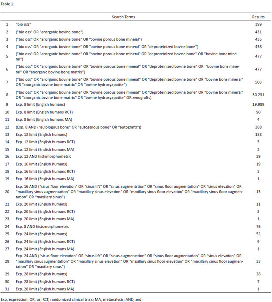
The database search strategy involved the use of MeSH terms and text words to create a specific (human, English, randomized controlled trials and meta-analysis) and sensitive database. The search strategy, did not established a defined time interval in order to reach a maximum number of evidences related to the subject.
The search terms used and results obtained in the PUBMED database are shown in Table 1.
Inclusion Criteria
All studies obtained through the search strategy containing maxillary sinus elevation with BioOss® xenogenous material associated or not with autogenous bone and that presented histomorphometric assessments. In addition, only studies in humans and published in English were considered.
As the number of randomized controlled trials and meta-analysis was too restricted, all levels of evidence were included in this review.
Studies with lack of data were also included in this review, to assess the quality of information published.
Exclusion Criteria
Studies involving multiple interventions (e.g., simultaneous alveolar ridge augmentation), studies with only histological data and abstracts.
RESULTS
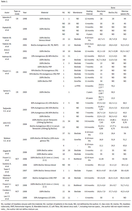
Thirty-nine studies involving histomorphometric analysis on maxillary sinuses elevation using xenogenous materials mixed or not with autogenous bone, written in English and performed in humans were identified. However, after applying the previously defined criteria, only 19 studies (12-30) were part of the scope of this review, and of these only three were randomized clinical trials (RCT) and none was a meta-analysis (MA). Of these 19 studies, six had a sample size too small, less than 10 patients (16, 22, 25, 27-29), six had no complete histomorphometric data such as neo-bone formation, remaining BioOss® particles and marrow spaces (12, 14, 17, 25-27), two lacked the sample size of grafted sinuses (12, 21), and six studies had no specific data such as the use of membranes, the proportion used in grafts composed by BioOss® and autogenous bone or even when using simple or composite grafts (12, 13, 15, 20, 22, 28). Thus, there were only five studies that contained a large enough sample size for possible analysis, complete histomorphometric data, definition healing time intervals and description of graft material used (18, 19, 23, 24, 30). Of these five, two were RCTs.
These five studies were elected to lead the objectives of this review. The results are shown in Table 2.
DISCUSSION
Results obtained through the studies contained in this review confirm the hypothesis that BioOss® has osteoconductive property. However, it is important to highlight the limitations and shortcomings of the studies that aimed to investigate these properties, for example, the methodological quality used, the sample size and inconsistent results presented, as we will discuss later.
New bone formation - The five studies which based this review provided, taking their limitations into account, sufficient data to demonstrate more clearly the hypothesis of BioOss® osteoconductive properties. Studies using different methods, but using the same type of graft (18, 23, 24) observed values ranging from 42.1% ± 10% to 14.7% ± 5% of neo-bone formation in healing time intervals ranging from 4 to 12 months. If we compare these values with the study by Froum, et al. (2008), where values ranging from 22.3% ± 6.4% were obtained at an interval of six to eight months, but using different BioOss® particle sizes, one can hypothesize that time is a determining factor for the amount of new bone that will form in maxillary sinuses grafted with only BioOss®. Some studies also corroborate this hypothesis (17, 25, 27), although the results presented in these studies were rather inconsistent or had very small populations.
However, because of the methodological drawbacks found by this study and the lack of specific control groups, such as remaining bone histomorphometry, it is not possible to conclude whether the values are somewhat good or bad, as all them except one (18), did not present histomorphometric comparisons with bone tissue from the patients' remaining alveolar ridge. According to Cordaro, et al. 2008, a low percentage of soft tissue graft was observed in their study, compared with bone tissue from the remaining alveolar ridge immediately below it, which allows us to hypothesize that even with supposedly low values, the BioOss® graft produces a denser trabecular bone than pre-existing bone, therefore providing excellent primary stability. As evidence of this, in a systematic review by Wallace et al., 2003, shows high implant survival rate in maxillary sinuses grafted with deproteinized bovine bone.
Due to the difficulty of creating control groups in procedures for maxillary sinuses elevation in humans, we suggest, as the work of Cordaro, et al. 2008, that ieces are obtained through an alveolar path, so that histomorphometric results can be compared between the graft and the remaining bone tissue immediately below, to determine the degree of biomaterial osteoconductivity.
Piattelli, et al. 1999, Scarano, et al. 1999, Scarano, et al. 2006 and Degidi, et al. 2006 found in their histomorphometric analyses a new bone formation composed of mature compact bone after a healing period of six months and using grafts made of 100% BioOss®, while Artzi, et al. 2001 using the same graft and extending the healing period to 12 months, find in their analysis a neo bone formation consisting of mainly young bone. Studies that used grafts associated with autogenous bone and similar healing times (14, 15, 26) found in their analysis a predominantly young bone formation combined with mature bone. There is a need for further studies to elucidate the processes of bone maturation in maxillary sinus elevation.
Association with autogenous bone - Eight papers were identified in this review using a mixture of BioOss® with autogenous bone in different proportions and featuring quite heterogeneous healing intervals. However, according to the analysis of the studies found and the variables required in this review, no studies provided the necessary information to hypothesize the benefits of exclusive addition of autogenous bone to grafts with BioOss®. However, using the investigated material (BioOss®) as a control group, heterogeneous healing time intra-study and a fairly small population, Tadjoedin, et al. 2003 demonstrate through their findings that the combination of autogenous bone and BioOss® in given proportions, increases the amount of neo bone formation, even extending the healing time of groups using grafts with smaller proportions of autogenous bone. They explain that autogenous bone particles mixed with the graft function as ossification centers, everywhere, by taking living cells and undifferentiated mesenchymal cells throughout the graft. Froum, et al. 2002 also found larger results of neo bone formation for the groups that received BioOss® grafts associated with autogenous bone when compared with the groups that received only BioOss® in a very similar period of healing. Hallman, et al. 2002, using the same mixing ratio and a similar healing period to that used by Tadjoedin, et al. 2003, found much higher values of neo bone formation, compared with the results presented by Tadjoedin, et al. 2003. In addition, still comparing these two studies, Hallman, et al. 2002 in their grafts with 100% BioOss® show results far superior to those found by Tadjoedin, et al. 2003.
However, due to methodological shortcomings of the analyzed studies, no conclusions can be drawn about the advantages of this association.
Association with PRP - According to the analysis of the studies found and the variables required in this review no studies provided the necessary information to hypothesize the benefits of associating BioOss® with PRP (Platelet Rich Plasma). However, the study by Froum et al. 2002, with a population of only three patients, using membranes with different properties between groups and no specific control groups, found values of 15% of neo bone formation for sinuses grafted with BioOss® and PRP and 13% when using only BioOss® on a healing period of seven months; and a value of 34% for sinus grafted with BioOss ® and PRP versus 32% without PRP, this time with a healing interval of 11 months. The author concludes that the addition of PRP to the graft does not generate statistically significant results both in the production of vital bone and the proportion of bone-implant contact, when compared with grafts without PRP. Comparing this with the result found in the study by Artzi, et al. 2001, it is possible to hypothesize that the addition of PRP does not produce increased neo bone formation when only BioOss® is used.
On the other hand, the authors of the studies included in this review that used PRP on the graft conclude that the use of PRP substantially improves graft handling.
However, it is worth noting the limitations of the studies found here and the need for large randomized clinical trials to assess the benefits and drawbacks of this association.
Association with autogenous and PRP - According to the analysis of the studies found and the variables required in this review only one study provided sufficient data to hypothesize the benefits of this association. According to GalindoMoreno, et al. 2007, using a composite graft of BioOss®, PRP and autogenous bone harvested with bone scraper from the bone wall of the maxillary sinus, the authors observed a neo bone formation by 34% ± 6.34%. When compared with studies that contained a mixture of BioOss® and autogenous bone, even in different proportions, but at similar healing intervals (13, 15, 20, 25, 28) the results obtained by Galindo-Moreno, et al. 2007 were consistently higher. However, the study by Hallman et al. 2002, found a value of 39.9% ± 8% for a mixture of 20% autogenous bone harvested from the mandibular branch and 80% BioOss® without the addition of PRP. Also in this same study, but with a slightly longer healing time than the study by Galindo-Moreno, et al. 2007 and using only BioOss® a value of 41.7% ± 26.6% was found. The results of the study by Artzi, et al. 2001 also resemble those found by Hallman, et al. 2002.
Thus nothing can be concluded about the association of PRP, BioOss® and autogenous bone, due to limitations of the studies and the enormous shortage of well-designed randomized clinical trials, so a hypothesis of the benefits of this triple combination can be formulated.
Remaining BioOss® particles - Among the works chosen to guide this review, two studies (19, 24) found lower amounts of remaining BioOss® particles in their analysis than neo bone formation, while Froum, et al. 2008, Cordaro, et al. 2008, and Yildirim et al. 2001 found values of remaining particles above the amount of neo bone formation. The values expressed here are not a determining factor for the analysis of graft quality, since, first, an ideal graft needs to maintain its dimensions stable over time and allow implant long term stability (31) thus needing be just a little reabsorbed or not at all, and second, there is a need to give a real definition of the true meaning of these particles within the graft as a whole. Since BioOss® is an osteoconductive material, it is assumed that it will guide and be incorporated by neo bone formation. In the studies by Lee et al. 2008, Piattelli, et al. 1999, Scarano, et al. 2006 and Degidi, et al. 2006 revitalization of Harvers Channels are reported in BioOss® particles. Fifteen studies (12-19, 21-24, 26, 27, 30), 19 of which will form the scope of this review, reported a close contact of BioOss® particles with new bone, which are partially or completely surrounded by it and osteoblasts deposit matrix bone directly onto its surface, confirming the antigenic and biocompatible properties of the material. The studies by GalindoMoreno, et al. 2007, Wallace, et al. 2005 and Traini, et al. 2007 reported interconnectivity between the particles through trabecular bridges of new bone, thus forming an integrated, robust and high-density graft, favoring the possibility of excellent primary stability of implants and corroborating its high rates of success.
Furthermore, in most studies analyzed, osteoclastic remodeling of BioOss® particles was observed, that for some authors (12, 13, 15-19, 21, 23, 27, 28) was considered slow, while for Lee, et al. 2008, Valentini, et al. 1998 and Hallman, et al. 2002 remodeling was absent. According Traini, et al. 2007, the hypothesis that could explain this slow absorption would be the high concentrations of Ca released in the medium due to acid secreted by osteoclasts on the particles, and by feedback, inhibiting them.
In Therefore, it seems beneficial to maintain BioOss® particles as part of the graft.
Marrow spaces – of the basic components of the marrow spaces, the presence of blood vessels for the occurrence of osteogenesis is undoubtedly the most importantLee, et al. 2008, Hallman, et al. 2002 and GalindoMoreno, et al. 2007 cite a rich network of vessels found in their studies and according to Degidi, et al. 2006, bone formation is closely related to the invasion of blood vessels, and they showed in their study a network of blood vessels located in the periphery of the marrow spaces intimately related to the new bone, but without observing vessels between BioOss® particles and new bone tissue. Nevertheless it is important to highlight the need for further studies to elucidate the angiogenic role of various biomaterials.
Traini, et al. 2007, Yildirim, et al. 2001, Piattelli, et al. 1999, Degidi, et al. 2006, Scarano, et al. 2006 and Hallman, et al. 2002 also report no inflammatory infiltrates, confirming the high biocompatibility of the material and its safe use.
CONCLUSION
With the limits of this systematic review, the following conclusions can be drawn:
1 - BioOss® has excellent osteoconductive properties;
2 - There are no sufficient scientific evidence to describe the benefits of associating BioOss® to autogenous bone. Randomized clinical trials should be conducted to elucidate this issue;
3 - The benefits of adding PRP to grafts in maxillary sinus elevation surgery has not yet been clarified;
4 - Remaining BioOss® particles provide high density grafts, favoring primary stability of implants, and it is beneficial to maintain them;
5 - Not enough evidence was found to statistically determine an average amount of standard neo bone formation, according to the protocols used and the methodology employed in the studies;
6 - With the available scientific evidence, it is not possible to obtain an ideal protocol to use BioOss® due to the heterogeneity of the studies and the results presented. Well-designed randomized trials should be conducted to elucidate this issue.
Future Directions
Clinical trials that limit the variables to evaluate a specific item are required to correctly identify and isolate the effects that are still considered confusing.
Clarification
The author has no direct or indirect financial interest in products mentioned in this article.
REFERENCES
1. Boyne PJ, James RA. Grafting of the maxillary sinus floor with autogenous marrow and bone. Journal of Oral Surgery. 1980 Aug;38(8):613-6.
2. Esposito M, Grusovin MG, Kwan S, Worthington HV, Coulthard P. Interventions for replacing missing teeth: bone augmentation techniques for dental implant treatment. Cochrane Database of Systematic Reviews (Online). 2008(3):CD003607.
3. Tatum H, Jr. Maxillary and sinus implant reconstructions. Dental Clinics of North America. 1986 Apr;30(2):207-29.
4. Barone A, Crespi R, Aldini NN, Fini M, Giardino R, Covani U. Maxillary sinus augmentation: histologic and histomorphometric analysis. The International Journal of Oral & Maxillofacial Implants. 2005 Jul-Aug;20(4):519-25.
5. Cawood JI, Stoelinga PJ. International academy for oral and facial rehabilitation--Consensus Report. International Journal of Oral and Maxillofacial Surgery. 2006 Mar;35(3):195-8.
6. Wallace SS, Froum SJ. Effect of maxillary sinus augmentation on the survival of endosseous dental implants. A systematic review. Annals of Periodontology. 2003 Dec;8(1):328-43.
7. Aghaloo TL, Moy PK. Which hard tissue augmentation techniques are the most successful in furnishing bony support for implant placement? The International Journal of Oral & Maxillofacial Implants. 2007;22 Suppl:49-70.
8. Crespi R, Vinci R, Cappare P, Gherlone E, Romanos GE. Calvarial versus iliac crest for autologous bone graft material for a sinus lift procedure: a histomorphometric study. The International Journal of Oral & Maxillofacial Implants. 2007 Jul-Aug;22(4):527-32.
9. Becktor JP, Hallstrom H, Isaksson S, Sennerby L. The use of particulate bone grafts from the mandible for maxillary sinus floor augmentation before placement of surface-modified implants: results from bone grafting to delivery of the final fixed prosthesis. Journal of Oral and Maxillofacial Surgery. 2008 Apr;66(4):780-6.
10. Silva FM, Cortez AL, Moreira RW, Mazzonetto R. Complications of intraoral donor site for bone grafting prior to implant placement. Implant Dentistry. 2006 Dec;15(4):420-6.
11. Cornell CN. Osteobiologics. Bulletin (Hospital for Joint Diseases (New York, NY)). 2004;62(1-2):13-7.
12. Piattelli M, Favero GA, Scarano A, Orsini G, Piattelli A. Bone reactions to anorganic bovine bone (Bio-Oss) used in sinus augmentation procedures: a histologic long-term report of 20 cases in humans. The International Journal of Oral & Maxillofacial Implants. 1999 Nov-Dec;14(6):835-40.
13. John HD, Wenz B. Histomorphometric analysis of natural bone mineral for maxillary sinus augmentation. The International Journal of Oral & Maxillofacial Implants. 2004 Mar-Apr;19(2):199-207.
14. Lee CY, Rohrer MD, Prasad HS. Immediate loading of the grafted maxillary sinus using platelet rich plasma and autogenous bone: a preliminary study with histologic and histomorphometric analysis. Implant Dentistry. 2008 Mar;17(1):59-73.
15. Yildirim M, Spiekermann H, Handt S, Edelhoff D. Maxillary sinus augmentation with the xenograft Bio-Oss and autogenous intraoral bone for qualitative improvement of the implant site: a histologic and histomorphometric clinical study in humans. The International Journal of Oral & Maxillofacial Implants. 2001 Jan-Feb;16(1):23-33.
16. Traini T, Valentini P, Iezzi G, Piattelli A. A histologic and histomorphometric evaluation of anorganic bovine bone retrieved 9 years after a sinus augmentation procedure. Journal of Periodontology. 2007 May;78(5):955-61.
17. Degidi M, Artese L, Rubini C, Perrotti V, Iezzi G, Piattelli A. Microvessel density and vascular endothelial growth factor expression in sinus augmentation using Bio-Oss. Oral Diseases. 2006 Sep;12(5):469-75.
18. Cordaro L, Bosshardt DD, Palattella P, Rao W, Serino G, Chiapasco M. Maxillary sinus grafting with Bio-Oss or Straumann Bone Ceramic: histomorphometric results from a randomized controlled multicenter clinical trial. Clinical Oral Implants Research 2008 Aug;19(8):796-803.
19. Galindo-Moreno P, Avila G, Fernandez-Barbero JE, Aguilar M, Sanchez-Fernandez E, Cutando A, et al. Evaluation of sinus floor elevation using a composite bone graft mixture. Clinical Oral Implants Research. 2007 Jun;18(3):376-82.
20. Wallace SS, Froum SJ, Cho SC, Elian N, Monteiro D, Kim BS, et al. Sinus augmentation utilizing anorganic bovine bone (Bio-Oss) with absorbable and nonabsorbable membranes placed over the lateral window: histomorphometric and clinical analyses. The International Journal of Periodontics & Restorative Dentistry. 2005 Dec;25(6):551-9.
21. Scarano A, Degidi M, Iezzi G, Pecora G, Piattelli M, Orsini G, et al. Maxillary sinus augmentation with different biomaterials: a comparative histologic and histomorphometric study in man. Implant Dentistry. 2006 Jun;15(2):197-207.
22. Valentini P, Abensur D, Densari D, Graziani JN, Hammerle C. Histological evaluation of Bio-Oss in a 2-stage sinus floor elevation and implantation procedure. A human case report. Clinical Oral Implants Research. 1998 Feb;9(1):59-64.
23. Yildirim M, Spiekermann H, Biesterfeld S, Edelhoff D. Maxillary sinus augmentation using xenogenic bone substitute material Bio-Oss in combination with venous blood. A histologic and histomorphometric study in humans. Clinical Oral Implants Research. 2000 Jun;11(3):217-29.
24. Artzi Z, Nemcovsky CE, Tal H, Dayan D. Histopathological morphometric evaluation of 2 different hydroxyapatite-bone derivatives in sinus augmentation procedures: a comparative study in humans. Journal of Periodontology. 2001 Jul;72(7):911-20.
25. Froum SJ, Wallace SS, Tarnow DP, Cho SC. Effect of platelet-rich plasma on bone growth and osseointegration in human maxillary sinus grafts: three bilateral case reports. The International Journal of Periodontics & Restorative Dentistry. 2002 Feb;22(1):45-53.
26. Hallman M, Sennerby L, Lundgren S. A clinical and histologic evaluation of implant integration in the posterior maxilla after sinus floor augmentation with autogenous bone, bovine hydroxyapatite, or a 20:80 mixture. The International Journal of Oral & Maxillofacial Implants. 2002 Sep-Oct;17(5):635-43.
27. Sartori S, Silvestri M, Forni F, Icaro Cornaglia A, Tesei P, Cattaneo V. Ten-year follow-up in a maxillary sinus augmentation using anorganic bovine bone (Bio-Oss). A case report with histomorphometric evaluation. Clinical Oral Implants Research. 2003;14:369-72.
28. Tadjoedin ES, de Lange GL, Bronckers AL, Lyaruu DM, Burger EH. Deproteinized cancellous bovine bone (Bio-Oss) as bone substitute for sinus floor elevation. A retrospective, histomorphometrical study of five cases. Journal of Clinical Periodontology. 2003 Mar;30(3):261-70.
29. Froum SJ, Wallace SS, Elian N, Cho SC, Tarnow DP. Comparison of mineralized cancellous bone allograft (Puros) and anorganic bovine bone matrix (Bio-Oss) for sinus augmentation: histomorphometry at 26 to 32 weeks after grafting. The International Journal of Periodontics & Restorative Dentistry. 2006 Dec;26(6):543-51.
30. Froum SJ, Wallace SS, Cho SC, Elian N, Tarnow DP. Histomorphometric comparison of a biphasic bone ceramic to anorganic bovine bone for sinus augmentation: 6- to 8-month postsurgical assessment of vital bone formation. A pilot study. The International Journal of Periodontics & Restorative Dentistry. 2008 Jun;28(3):273-81.
31. Block MS, Kent JN. Sinus Augmentation for Dental Implants: The Use of Autogenous Bone. Journal of Oral and Maxillofacial Surgery. 1997;55:1281-6.
1. Specialist Dentist. 2. Master's degree Dentist. 3. Master's degree Dentist. 4. Master's degree Dentist.
Correspondence: João Surerus de Oliveira Junior Rua Oscar Vidal, 71, sala 603, Centro Juiz de Fora MG, Brazil Phone/Fax: 55 032 30613016 Email: jsreabilitacaooral@hotmail.com
This article was submitted to SGP (Publication Management System) of Rev Bras Cien Med Health on March 7, 2010. Cod. 53. Accepted on May 13, 2010.