
INTRODUCTION
Bloodstream infections (BSI) are related to unfavorable health outcomes. In Brazil, through the Brazilian SCOOPE study, about 40% of mortality was observed among patients with BSI1. The presence of bacteria or fungi in the bloodstream is called bacteremia and fungemia, respectively2. Intravascular devices, genitourinary tract, respiratory tract, intestine, biliary tract, intra-abdominal abscess, and unknown sites are the most common sources of BSI3,4.
The body's response to the infectious agent occurs through signs and symptoms of the disease, such as the systemic inflammatory response syndrome, regardless of whether blood culture is positive or not. This set of responses is called sepsis. This, in turn, promotes organic dysfunction and puts the patient's life at risk. Despite the precariousness of studies on incidence and prevalence related to sepsis in Brazil, it is known that this syndrome is considered a major public health problem in intensive care units (ICUs), generating high costs for health systems3,5, 6.
The blood culture test is the gold standard for the detection of microorganisms in the blood. This has a high predictive value of infection since it is possible to detect the presence of viable microorganisms in the bloodstream. Its positivity is a relevant indicator of BSI and the identification of the causative pathogen is of utmost importance since it helps in the therapeutic choice3,5. Therefore, inadequate therapeutic regimens induce selective pressure, which results in increased resistant strains, increased hospitalization costs, and higher mortality rates, especially in critically ill patients6, 7, 8,9.
In view of the above, the aim of the present study was to assess the prevalence of isolated microorganisms in blood samples from patients in the ICUs and Coronary Care Unit (CCU) of a private hospital in the city of Juiz de Fora, Minas Gerais, and to evaluate the susceptibility profile to the antimicrobials of the isolated bacterial strains, thus contributing to research in the area of prevention of BSI, in addition to encouraging the Rational Use of Medicines.
METHODS
This is a descriptive, cross-sectional retrospective study and aimed to analyze blood culture samples performed in the clinical analysis laboratory of a private hospital located in the city of Juiz de Fora, Minas Gerais, from January 2017 to January 2019 .
As an inclusion criterion, all samples of positive blood cultures were considered. The data that are part of the research were obtained through the digital files of the laboratory software Shift Lis® used by the laboratory. As this is a statistical analysis of data from exams already performed, this research represents minimal risks to patients, but significant in relation to the manipulation of digital data.
This study was previously approved by the Research Ethics Committee in accordance with Resolution 196/96 of the National Health Council under Opinion number 3.735.713.
The included blood cultures were automatically detected by the BACT/ALERT® 3D 60 device from bioMérieux, with subsequent identification and antibiogram in the Vitek® 2 Compact system from bioMérieux with GN identification cards for Gram-negative fermenters and non-fermenters with antibiogram cards AST-N238 and AST-N239, GP for Gram-positive and its complementary antibiogram card AST-P585, in addition to the YST for yeast identification10.
RESULTS
A total of 3536 blood culture samples were collected from the ICUs and CCUs over a two-year period, 2590 (73.3%) from the Adult ICU, 540 (15.3%) from the Neonatal ICU and 406 (11.5%) from the CCU. Of the total samples collected, 1072 (30.3%) showed positive growth and included the present study.
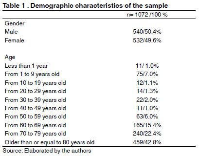
According to Table 1, among the samples analyzed, 540 (50.4%) were male patients, while 532 (49.6%) were female. It was also observed that more than 80% of the patients were 60 years old or older.
In the Adult ICU, 865 samples were positive, which correspond to 80.7% of the total samples analyzed. The rest of the samples came from CCU (113/10.5%) and Neonatal ICU (94/8.8%).
Of the 1072 samples studied, 44 showed concomitant growth of two microorganisms, totaling 1116 isolated microorganisms. Of these, 704 (63.1%) were Gram-positive cocci, 327 (29.3%) were morphotintorially classified as Gram-negative rods, 58 (5.2%) presented as fungal forms and 27 (2.4%) like Gram-positive rods.
According to Table 2, the prevalent microorganisms were: Staphylococcus sp.coagulase-negative (55.5%), Klebsiella pneumoniae (7.2%), Staphylococcus aureus (5.4%), Serratia marcescens (4.7 %), Escherichia coli (3.9%), Acinetobacter baumannii (3.6%), Pseudomonas aeruginosa (2.4%), Gram- positive rods (2.4%), Candida parapsilosis (2.2%), Enterobacter cloacae (2.0%), Enterococcus faecalis (1.4%) and Candida albicans (1.3%).
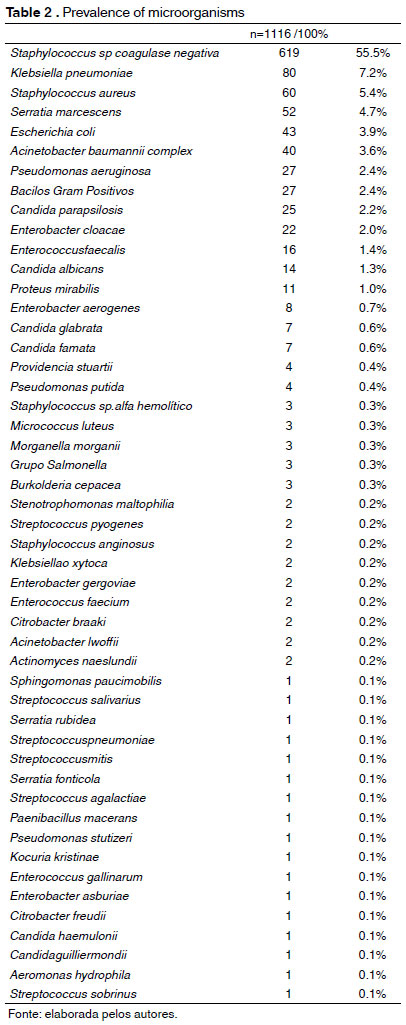
The isolates belonging to the genus Staphylococcus sp.were prevalent (666/59.7%), followed by the order Enterobacteriales (237/21.2%), Gram- negative non-fermenting rods (78/7.0%), fungi (57/5.1%), Gram-positive bacilli (27/2.4%), and Gram-positive cocci of the genus Enterococcus sp. (19/1.7%) as described in Table 2.
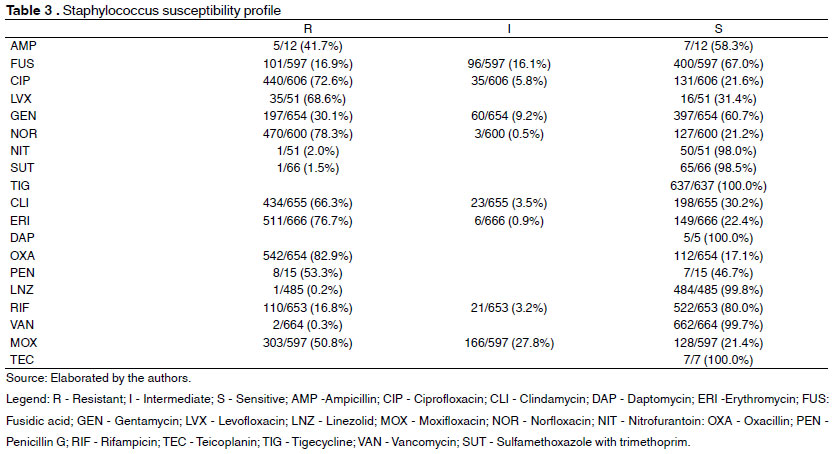
Among staphylococci, in Table 3 we have their sensitivity profile, where the greatest resistance found was oxacillin (82.9%), followed by erythromycin (76.7%). As for quinolones, 78.3% of staphylococci were resistant to norfloxacin, 72.6% to ciprofloxacin, 68.6% to levofloxacin and 50.8% to moxifloxacin.
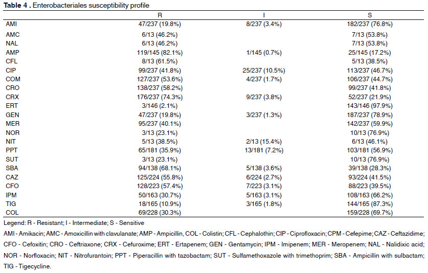
In the susceptibility test to antimicrobials, microorganisms belonging to the order Enterobacteriales had a high resistance index. Among cephalosporins, 74.3% showed resistance to cefuroxime, 61.5% to cephalothin, 58.2% to ceftriaxone, 55.8% to ceftazidime, and 53.6% to cefepime. Regarding quinolones, 46.2% of enterobacteria showed resistance to nalidixic acid, 41.8% to ciprofloxacin, and 23.1% to norfloxacin; 82.1% of the strains were resistant to ampicillin and 68.1% to ampicillin with sulbactam; 23.1% were resistant to sulfamethoxazole with trimethoprim; 19.8% of enterobacteria were resistant to amikacin and gentamicin. It was also observed a high resistance of these enterobacteria to carbapenems, where 40.1% were resistant to meropenem, 30.7% to imipenem, and 2.1% to ertapenem. Finally, 57.4% were resistant to cefoxitin, 38.5% to nitrofurantoin, 35.9% to piperacillin with tazobactam, 30.3% to colistin, and 10.9% to tigecycline. Such data were described in Table 4.
Of the 237 isolated enterobacteria, 14.4% (34/237) had a beta-lactamase resistance mechanism of the extended-spectrum (ESBL). Of these, 73.5% (25/34) were K. pneumoniae and 26.5% (9/34) E.coli.
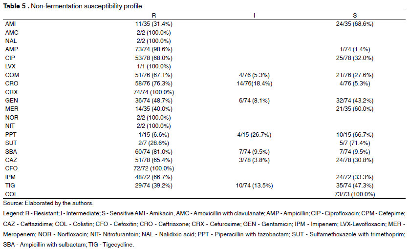
In the analysis of the antimicrobial susceptibility profile of non-fermenting Gram-negative rods, a high percentage of resistance was observed. Among cephalosporins, 100% of the strains analyzed were resistant to cefuroxime, 76.3% to ceftriaxone, 67.1% to cefepime, and 65.4% to ceftazidime. As for quinolones, 100% of the non-fermenters were resistant to nalidixic acid, levofloxacin, and norfloxacin, while 68% were resistant to ciprofloxacin. Regarding aminoglycosides, 48.7% of the bacteria showed resistance to gentamicin and 31.4% to amikacin. Among the carbapenems, 66.7% were resistant to imipenem and 40% to meropenem. 100% of the strains were resistant to nitrofurantoin, cefoxitin, and amoxicillin with clavulanic acid. The resistance of these non-fermenters extends to tigecycline and piperacillin with tazobactam with 39.2% and 6.6% resistance, respectively, according to Table 5.
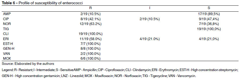
Among the 19 strains of isolated enterococci, 100% showed resistance to clindamycin, streptomycin, and high-concentration gentamicin. Among quinolones, it was observed that 100% of the bacteria were resistant to moxifloxacin, 63.2% to norfloxacin, and 42.1% to ciprofloxacin. In addition to these antimicrobials, some strains were also tested for vancomycin and ampicillin, with 100% of them showing resistance to vancomycin and 10.5% to ampicillin, as described in Table 6.
DISCUSSION
In a study by Rodriguéz et al. (2011) 1045 blood cultures of clinical interest were analyzed, of which 375 (35%) were positive for some pathogen and 670 (65%) were negative. According to Chen et al. (2015) and Weiss et al. (2016), the proportionality of positivity of blood cultures remains, where the rate of positivity varies between 10% and 30%. Thus, it is possible to state that the results obtained in the present study were compatible: Of the total of 3536 blood culture samples, 2464 (69.7%) had a negative result, while 1072 (30.3%) were positive for the growth of some microorganism.
The present study showed that the positive blood cultures analyzed had a higher incidence in males 540 (50.7%) compared to females 532 (49.3%). Corroborating this result is the study performed by Sousa et al (2014) in which test results from 170 positive blood cultures of patients hospitalized in the ICU were evaluated, where about 56.0% were male and 43.0% of women. According to Couto et al (2011), this is probably due to the fact that female hormones, especially estrogen, promote a better immune response to women. In addition, Wang et al. (2015) showed that about 60.0% of severe sepsis occurred in patients over 60 years of age, calling attention to the high percentage of elderly people with positive blood cultures found in this study.
According to Morello et al. (2019) and Takeshita et al. (2019) coagulase-negative staphylococci were the most commonly isolated pathogens and the high prevalence of these can be confirmed during the study, with the isolation of 619 species, representing 55.46% of the total pathogens isolated from blood cultures. According to Park et al. (2015), coagulase-negative staphylococci are responsible for 20 to 30% of infections in critically ill patients and are generally associated with central catheters and implantable devices. The high prevalence of these microorganisms indicates a possible contamination at the time of collection, since this pathogen can be found in several sites on the body, including the composition of the skin microbiota. However, due to the retrospective nature of this study, it was not possible to assess whether the detection of this pathogen was due to infection or contamination of the samples.
As for the profile of susceptibility to antimicrobials, a study by Ruschel et al. (2017) found high staphylococcal resistance to oxacillin, erythromycin, clindamycin, and quinolones, corroborating the research.
The main Gram-negative bacilli found in the present study were Klebsiella pneumoniae, Serratia marcescens, Escherichia coli and Enterobacter cloacae. This prevalence was similar to that reported by Basso et al. (2016), Mota et al. (2018), and Uc-Cachónet al (2019), where among the main pathogens classified as Gram-negative bacilli were Klebsiella pneumoniae and Escherichia coli.
The high rate of resistance of enterobacteria to cephalosporins, quinolones, aminoglycosides, and carbapenems described in this study are similar to those reported in a study performed in a teaching hospital by Parajuli et al. (2017) and according to UC-Cachón et al. (2019), the high rate of resistance to cephalosporins is confirmed.
As for the production of ESBL, the study showed a low percentage (14.4%), differing from the study by Myat et al. (2017), who found about 38%. However, both studies found a prevalence of Klebsiella pneumoniae among strains producing extended-spectrum beta-lactamase, followed by Escherichia coli.
The prevalence found of non-fermenting Gram-negative rods was Acinetobacter baumannii and Pseudomonas aeruginosa, the same as reported by Basso et al. (2016), Mota et al. (2018). In addition, the high rate of resistance to quinolones, cephalosporins, aminoglycosides, carbapenems described in this study, is similar to that described by Moolchandani et al. (2017), where the non-fermenters isolated by them showed resistance to the same classes of antimicrobials.
When it comes to fungemia, it is noted that the main associated fungus is Candida albicans (19,21,22,23). In Brazil, the survey of epidemiological data on fungemias in communities and hospitals has become increasingly frequent. In their study, Motta et al. (2010), reported a prevalence of 4% of fungemia in blood cultures with approximately 86% of cases related to species of the genus Candida sp. However, according to Silva et al. (2015), the current scenario has been showing an increase in the incidence of candidemia caused by non-albicans species, mainly by Candida parapsilosis. Such studies are in agreement with the present study since it was observed a prevalence of about 5% of forms fungi in the positive blood cultures analyzed, with a greater frequency of the species Candida parapsilosis and Candida albicans.
According to Arias et al. (2010) the most effective lactam beta against infections caused by enterococci is ampicillin, corroborating the study since this antimicrobial showed 89.5% sensitivity. High sensitivity to tigecycline is also noted, reaffirming the choice of this drug as a treatment alternative. However, Arias et al. warn that the prescription of such a drug should be done with caution since cases of resistance have already been reported.
CONCLUSION
Therefore, it was noted that the prevalence of negative coagulase Staphylococcus was prevalent, which serves as a warning for a possible infection caused by this microorganism, and should be analyzed together with the patient's clinic, since, according to the current literature, they are responsible for contamination of blood cultures, resulting from collection errors. In order to discover the real etiology of these infections, it is necessary to emphasize the correct asepsis of the professionals' hands in order to maintain patient safety and quality of the final result of the sample.
The study revealed a relevant prevalence of fermenting and non-fermenting enterobacteria with a high resistance profile to most of the antimicrobials used in medical practice, which encourages the rational use of medications.
New drugs and new control measures are needed, given that this growing resistance has become a worldwide public health problem.
REFERENCES
1. Agência Nacional de Vigilância Sanitária. Medidas de Prevenção de Infecção Relacionada à Assistência à Saúde. Brasília: Agência Nacional de Vigilância Sanitária, 2010.
2. Castro LL, Schutze M, Bucker DH, Vasconcellos LS. Prevalence of fungemia in a tertiary hospital: Analysis of the last decade. Rev Assoc Med Bras. 2016; 62(4): 315-319.
3. Araujo MRE. Hemocultura: recomendações de coleta, processamento e interpretação de resultados. J Infect Control. 2012; 1(1): 08-19.
4. Klingspor L, Ullberg M, Rydberg J, Kondori N, Serrander L, Swanberg J et al.Epidemiology of fungaemia in Sweden: A nationwide retrospective observational survey.Mycoses. 2018; 6: 777-785.
5. Ferreira LE, Dalposso K, Hackbarth BB, Gonçalves AR, Westphal GA, França PHC at al. Painel molecular para detecção de microrganismos associados à sepse. RevBras Ter Intensiva. 2011; 23(1): 36-40.
6. Sitnik R, Marra AR, Petroni RC, Ramos OPS, Martino MDV, Pasternak Jat al. Uso do SeptiFast para diagnóstico de sepse em doentes graves de um hospital brasileiro. Einstein. 2014; 12(2): 191-7.
7. Sociedade Brasileira de Patologia Clínica. Recomendações da Sociedade Brasileira de Patologia Clínica/Medicina Laboratorial (SBPC/ML):Boas práticas em microbiologia clínica. Barueri: Sociedade Brasileira de Patologia Clínica, 2015.
8. Siqueira JPZ, Almeida MAG. Molecular biology as a tool for fungal detection on blood: diagnostic aid and cost reduction. Arch. Helth. Sci. 2018; 25(3):41-45.
9. Giolo MP, Svidzinski TIE. Fisiopatogenia, epidemiologia e diagnóstico laboratorial da candidemia. J BrasPatolMed Lab. 2010; 46:225-34.
10. bioMérieux Brasil. Produtos e Serviços. Disponível em: URL: http://www.biomerieux.com.br/diagnostico-clinico/produtos-e-servicos. Acesso em 3 dezembro 2019.
11. Basso ME, Pulcinellii RSR, Aquino ARC, Santos KF. Prevalência de infecções bacterianas em pacientes internados em uma unidade de terapia intensiva (UTI). RevBras Anal Clin. 2016; 48(4): 383-88.
12. Mota FS, Oliveira HÁ, Souto RCF. Perfil e prevalência de resistência aos antimicrobianos de bactérias Gram-negativas isoladas de pacientes de uma unidade de terapia intensiva. Rev Bras Anal Clin. 2018; 50(3): 270-2.
13. Weiss SL, Fitzgerald JC, Pappachan J, Wheeler D, Jaramillo-Bustamante JC, Salloo A, Singhi SC, Erickson S, Roy JA, Bush JL, Nadkarni VM, Thomas NJ; Sepsis Prevalence, Outcomes, and Therapies (SPROUT) Study Investigators and Pediatric Acute Lung Injury and Sepsis Investigators (PALISI) Network. Global epidemiology of pediatric severe sepsis: the sepsis prevalence, outcomes, and therapies study. Am J RespirCrit Care Med. 2015; 191(10):1147-57. Erratum in: Am J RespirCrit Care Med. 2016; 193(2):223-4.
14. Chen XC, Yang YF, Wang R, Gou HF, Chen XZ. Epidemiology and microbiology of sepsis in mainland China in the first decade of the 21st century.Int J Infect Dis. 2015; 3: 9-14.
15. Park SY, Kwon KH, Chung JW, Huh HJ, Chae SL. Coagulase-negative staphylococcal bacteremia: risk factors for mortality and impact of initial appropriate antimicrobial therapy on outcome. Eur J ClinMicrobiolInfectDis. 2015; 34(7):1395-401.
16. Rodriguez F, Barrera L, De La Rosa G, Dennis R, Duenas C, Granados M, et al. The epidemiology of sepsis in Colombia: a prospective multicenter cohort study in tem university hospitals. Crit Care Med. 2011;39(7):1675-82.
17. Uc-Cachón AH, Osorno CG, Luna-Chi IG, Guillermo JGJ, Salinas GMM. High Prevalence of Antimicrobial Resistance AmongGram-Negative Isolated Bacilli in Intensive Care Units at a Tertiary-Care Hospital in Yucatán Mexico.Medicina.2019, 55, 588.
18. Morello LG, Dalla-Costa LM, Fontana RM, Netto ACSO, Petterle RR, Conte D, Pereira LA, Krieger MA, Raboni SM. Avaliação das características clínicas e epidemiológicas de pacientes com e sem sepse nas unidades de terapia intensiva de um hospital terciário. Einstein (São Paulo). 2019; 17(2): 1-8.
19. Anunnatsiri S, Chetchotisakd P, Mootsikapun P. Fungemia em pacientes não infectados pelo HIV: uma revisão de cinco anos. Int J InfectDis. 2009; 13 (1): 90-6.
20. Motta AL, Almeida DGM, Almeida Júnior JN, Burattini MN, Rossi, F. Candidemia epidemiologia e susceptibilidade perfil na maior complexo hospital universitário brasileiro. Braz J InfectDis. 2010; 14 (5): 441-8.
21. Colombo AL, Braga T, Silva LR, de Almeida Monfardini LP, Cunha AK, Rady P, et al. estudo prospectivo observacional de candidemia em São Paulo, Brasil: taxa de incidência, epidemiologia e preditores de mortalidade. Infect Controlo HospEpidemiol. 2007; 28 (5): 570-6.
22. Almirante B, D Rodriguez, Parque BJ, Cuenca-Estrella H, aplanam SOU, Almela H, et al .; Barcelona Candidemia Projeto StudyGroup. Epidemiologia e preditores de mortalidade em casos de infecção de corrente sanguínea Candida: resultados de vigilância de base populacional, Barcelona, Espanha, de 2002 a 2003. J Clin Microbiol. 2005; 43 (4): 1829-1835.
23. Costa SF, Marinho I, Araújo EA, Manrique AE, Medeiros EA, Levin AS. fungemianosocomial: um estudo prospectivo de 2 anos. J Hosp Infect. 2000; 45 (1): 69-72.
24. Myat TO, Hannaway RF, Zin KN, Htike WW, Win KK, Crump JA, Murdoch DR, Ussher JE. ESBL- and Carbapenemase-Producing Enterobacteriaceae in Patients with Bacteremia, Yangon, Myanmar, 2014.EmergingInfectiousDiseases. 2017; 23(5): 857-859.
25. Couto DO, Junior AAP, Farias JLM, Sales DB, Lima JPA, Rodrigues RS, Meneses FA. Associação entre sexo e mortalidade em pacientes com sepse: os hormônios sexuais influenciam o desfecho?. Revista brasileira de Terapia Intensiva. 2011; 23 (3): 297-303.
26. Wang HE, Donelly JP, Shapiro NI, Hohmann SF, Levitan EB. Hospital variations in severe sepsis mortality.Am J Med Qual. 2015; 30 (4): 328-336.
27. Sousa MA, Medeiros NM, Carneiro JR, Cardosos AM. Hemoculturas positivas de pacientes da unidade de terapia intensiva de um hospital escola de Goiânia, GO, entre 2010 e 2013. Estudos, Goiânia. 2014; 41(3): 627-635.
28. TakeshitaN, QuocAnh N, Phuong VT, Thuy, PP, Houng MTL, Takahashi, M and Ohmagari, N. Assessment of Bacteremia in a Large Tertiary Care Hospital in Northern Vietnam:a Single-Center Retrospective Surveillance Study. Jpn. J. Infect. Dis. 2019; 72: 118-120.
29. Silva MGC, Rodrigues GS, Gonçalves IL, Grazziotin NA. Distribuição de espécies de Candida e suscetibilidade ao fluconazol de isolados de hemocultivos em hospital regional de Passo Fundo, RS, Brasil. J. Bras. Patol. Med. Lab.2015; 51(3): 158-161.
30. Parajuli NP, Acharya SP, Mishra SK, Parajuli K. Basista Prasad Rijal and Bharat Mani PokhrelHigh burden of antimicrobial resistance among gram negative bacteria causing healthcare associated infections in a critical care unit of Nepal. Antimicrobial Resistance and Infection Control.2017; 6:67.
31. Moolchandani K, Sastry AS, Deepashree R, Sisltla S, Harish BM, Mandal J. Antimicrobial Resistance Surveillance among Intensive Care Units of a Tertiary Care Hospital in Southern India. Journal of Clinical and Diagnostic Research.2017; 11(2): 1-7.
32. Ruschel DB, Rodrigues AD, Formolo F. Perfil de resultados de hemoculturas positivas e fatores associados. RBAC. 2017; 49(2): 158-63.
33. Arias CA, Contreras GA, Murray BE. Management of multi drug-resistant enterococcal infections. Clin. Microbiol. Infect. 2010; 16: 555-562.